
Radiology Illustrated – serie
Visar alla böcker i serien Radiology Illustrated. Handla med fri frakt och snabb leverans.
15 produkter
15 produkter
Inbunden, Engelska, 2011
3 885 kr
Skickas inom 5-8 vardagar
Uroradiology is an up-to-date, image-oriented reference in the style of a teaching file that has been designed specifically to be of value in clinical practice. All aspects of the imaging of urologic diseases are covered, and case studies illustrate the findings obtained with the relevant imaging modalities in both common and uncommon conditions. Most chapters focus on a particular clinical problem, but normal findings, congenital anomalies, and interventions are also discussed and illustrated.In this second edition, the range and quality of the illustrations have been enhanced, and many schematic drawings have been added to help readers memorize characteristic imaging findings through pattern recognition. The accompanying text is concise and informative. Besides serving as an outstanding aid to differential diagnosis, this book will provide a user-friendly review tool for certification or recertification in radiology.
Inbunden, Engelska, 2012
3 663 kr
Skickas inom 5-8 vardagar
Radiology Illustrated: Gynecologic Imaging is an up-to-date, image-oriented reference in the style of a teaching file that has been designed specifically to be of value in clinical practice. Individual chapters focus on the various imaging techniques, normal variants and congenital anomalies, and the full range of pathology. Each chapter starts with a concise overview, and abundant examples of the imaging findings are then presented. In this second edition, the range and quality of the illustrations have been enhanced, and image quality is excellent throughout. Many schematic drawings have been added to help readers memorize characteristic imaging findings through pattern recognition. The organization of chapters by disease entity will enable readers quickly to find the information they seek. Besides serving as an outstanding aid to differential diagnosis, this book will provide a user-friendly review tool for certification or recertification in radiology.
Inbunden, Engelska, 2014
3 885 kr
Skickas inom 10-15 vardagar
This case-based atlas presents images depicting the findings typically observed when imaging a variety of common and uncommon diseases in the pediatric age group. The cases are organized according to anatomic region, covering disorders of the brain, spinal cord, head and neck, chest, cardiovascular system, gastrointestinal system, genitourinary system, and musculoskeletal system. Cases are presented in a form resembling teaching files, and the images are accompanied by concise informative text. The goal is to provide a diagnostic reference suitable for use in daily routine by both practicing radiologists and radiology residents or fellows. The atlas will also serve as a teaching aide and a study resource, and will offer pediatricians and surgeons guidance on the clinical applications of pediatric imaging.
Inbunden, Engelska, 2014
2 777 kr
Skickas inom 10-15 vardagar
Radiology Illustrated: Gastrointestinal Tract is the second of two volumes designed to provide clear and practical guidance on the diagnostic imaging of abdominal diseases. The book presents approximately 300 cases with 1500 carefully selected and categorized illustrations of gastrointestinal tract diseases, along with key text messages and tables that will help the reader easily to recall the relevant images as an aid to differential diagnosis. Essential points are summarized at the end of each text message to facilitate rapid review and learning. Additionally, brief descriptions of each clinical problem are provided, followed by case studies of both common and uncommon pathologies that illustrate the roles of the different imaging modalities, including ultrasound, radiography, computed tomography, and magnetic resonance imaging.
Häftad, Engelska, 2017
2 655 kr
Skickas inom 10-15 vardagar
Radiology Illustrated: Gynecologic Imaging is an up-to-date, image-oriented reference in the style of a teaching file that has been designed specifically to be of value in clinical practice. Individual chapters focus on the various imaging techniques, normal variants and congenital anomalies, and the full range of pathology. Each chapter starts with a concise overview, and abundant examples of the imaging findings are then presented. In this second edition, the range and quality of the illustrations have been enhanced, and image quality is excellent throughout. Many schematic drawings have been added to help readers memorize characteristic imaging findings through pattern recognition. The organization of chapters by disease entity will enable readers quickly to find the information they seek. Besides serving as an outstanding aid to differential diagnosis, this book will provide a user-friendly review tool for certification or recertification in radiology.
Häftad, Engelska, 2016
2 766 kr
Skickas inom 10-15 vardagar
This case-based atlas presents images depicting the findings typically observed when imaging a variety of common and uncommon diseases in the pediatric age group. The cases are organized according to anatomic region, covering disorders of the brain, spinal cord, head and neck, chest, cardiovascular system, gastrointestinal system, genitourinary system, and musculoskeletal system. Cases are presented in a form resembling teaching files, and the images are accompanied by concise informative text. The goal is to provide a diagnostic reference suitable for use in daily routine by both practicing radiologists and radiology residents or fellows. The atlas will also serve as a teaching aide and a study resource, and will offer pediatricians and surgeons guidance on the clinical applications of pediatric imaging.
Häftad, Engelska, 2016
2 001 kr
Skickas inom 10-15 vardagar
Radiology Illustrated: Gastrointestinal Tract is the second of two volumes designed to provide clear and practical guidance on the diagnostic imaging of abdominal diseases. The book presents approximately 300 cases with 1500 carefully selected and categorized illustrations of gastrointestinal tract diseases, along with key text messages and tables that will help the reader easily to recall the relevant images as an aid to differential diagnosis. Essential points are summarized at the end of each text message to facilitate rapid review and learning. Additionally, brief descriptions of each clinical problem are provided, followed by case studies of both common and uncommon pathologies that illustrate the roles of the different imaging modalities, including ultrasound, radiography, computed tomography, and magnetic resonance imaging.
Häftad, Engelska, 2016
2 125 kr
Skickas inom 11-20 vardagar
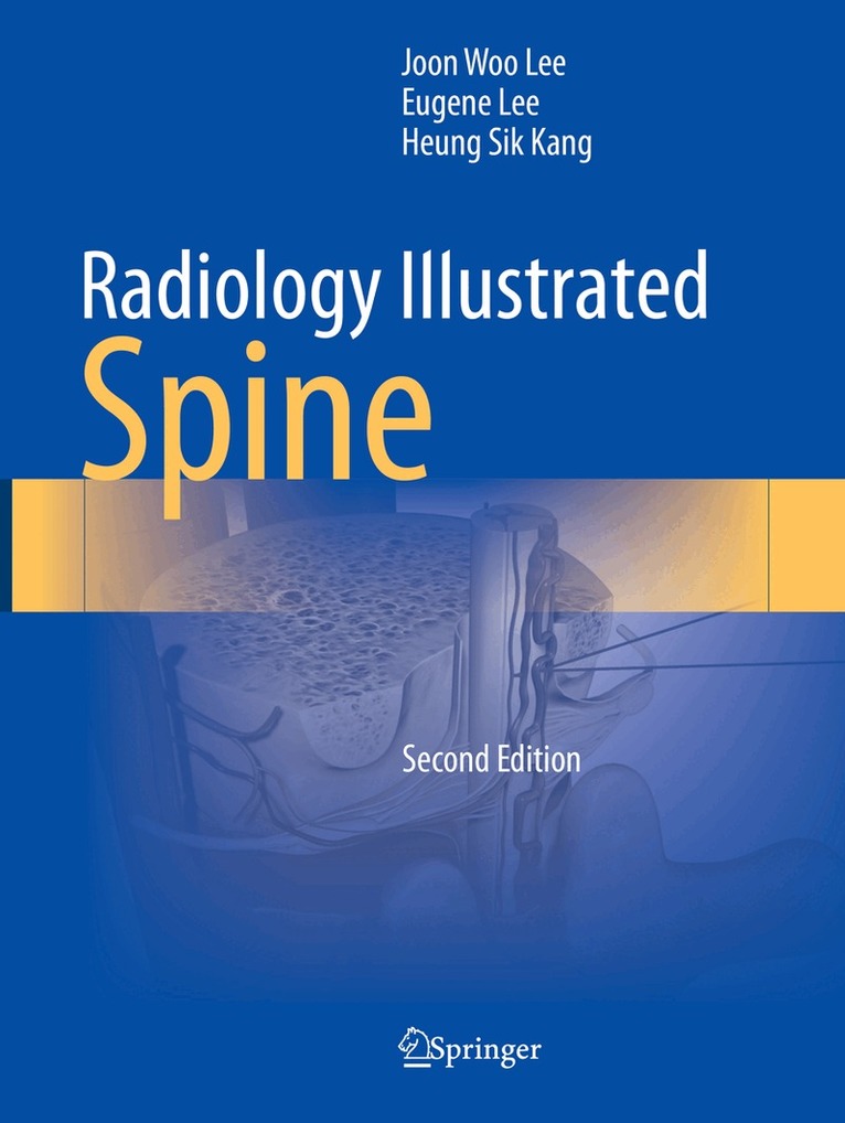
Radiology Illustrated: Spine is an up-to-date, superbly illustrated reference in the style of a teaching file that has been designed specifically to be of value in clinical practice. Common, critical, and rare but distinctive spinal disorders are described succinctly with the aid of images highlighting important features and informative schematic illustrations. The first part of the book, on common spinal disorders, is for radiology residents and other clinicians who are embarking on the interpretation of spinal images. A range of key disorders are then presented, including infectious spondylitis, cervical trauma, spinal cord disorders, spinal tumors, congenital disorders, uncommon degenerative disorders, inflammatory arthritides, and vascular malformations. The third part is devoted to rare but clinically significant spinal disorders with characteristic imaging features, and the book closes by presenting practical tips that will assist in the interpretation of confusing cases.
Häftad, Engelska, 2016
2 766 kr
Skickas inom 10-15 vardagar
Radiology Illustrated: Hepatobiliary and Pancreatic Radiology is the first of two volumes that will serve as a clear, practical guide to the diagnostic imaging of abdominal diseases. This volume, devoted to diseases of the liver, biliary tree, gallbladder, pancreas, and spleen, covers congenital disorders, vascular diseases, benign and malignant tumors, and infectious conditions. Liver transplantation, evaluation of the therapeutic response of hepatocellular carcinoma, trauma, and post-treatment complications are also addressed.The book presents approximately 560 cases with more than 2100 carefully selected and categorized illustrations, along with key text messages and tables, that will allow the reader easily to recall the relevant images as an aid to differential diagnosis. At the end of each text message, key points are summarized to facilitate rapid review and learning. In addition, brief descriptions of each clinical problem are provided, followed by both common and uncommon case studies that illustrate the role of different imaging modalities, such as ultrasound, radiography, CT, and MRI.
Inbunden, Engelska, 2022
1 669 kr
Skickas inom 10-15 vardagar
The nutcracker phenomenon (NCP) is an accentuated phenomenon of normal subtle compression of the left renal vein (LRV) between the abdominal aorta and superior mesenteric artery. The nutcracker syndrome (NCS) is a syndrome caused by NCP, resulting in hematuria, proteinuria, or left flank pain. The established diagnostic criterion of NCP is a pressure gradient more than 3 mmHG across the compression site, and NCS has been known to be rare, but probably is not rare and in fact quite common. The most important reason why NCS is known to be rare is that the diagnosis is difficult unless invasive venous catheterization and pressure measurement confirm the pressure gradient. Doppler ultrasound is useful in measuring the flow velocity of LRV. From the velocity measured by Doppler ultrasound, we may estimate the pressure gradient. Doppler ultrasound of LRV needs technical skills and experience. Computed tomography (CT) is a popular imaging technique for patients with hematuria, and we need to be familiar with the findings of the NCP at CT. There are variations of NCP associated with vascular anatomy and posture of the patients. This book will illustrate the concepts of NCP and NCS, findings at Doppler ultrasound and CT, and variations of NCP.
Häftad, Engelska, 2023
1 226 kr
Skickas inom 10-15 vardagar
The nutcracker phenomenon (NCP) is an accentuated phenomenon of normal subtle compression of the left renal vein (LRV) between the abdominal aorta and superior mesenteric artery. The nutcracker syndrome (NCS) is a syndrome caused by NCP, resulting in hematuria, proteinuria, or left flank pain. The established diagnostic criterion of NCP is a pressure gradient more than 3 mmHG across the compression site, and NCS has been known to be rare, but probably is not rare and in fact quite common. The most important reason why NCS is known to be rare is that the diagnosis is difficult unless invasive venous catheterization and pressure measurement confirm the pressure gradient. Doppler ultrasound is useful in measuring the flow velocity of LRV. From the velocity measured by Doppler ultrasound, we may estimate the pressure gradient. Doppler ultrasound of LRV needs technical skills and experience. Computed tomography (CT) is a popular imaging technique for patients with hematuria, and we need to be familiar with the findings of the NCP at CT. There are variations of NCP associated with vascular anatomy and posture of the patients. This book will illustrate the concepts of NCP and NCS, findings at Doppler ultrasound and CT, and variations of NCP.
Inbunden, Engelska, 2023
3 331 kr
Skickas inom 10-15 vardagar
Radiology Illustrated: Spine is an up-to-date, superbly illustrated reference in the style of a teaching file that has been designed specifically to be of value in clinical practice. Common, critical, and rare but distinctive spinal disorders are described succinctly with the aid of images highlighting important features and informative schematic illustrations. The first part of the book, on common spinal disorders, is for radiology residents and other clinicians who are embarking on the interpretation of spinal images. A range of key disorders are then presented, including infectious spondylitis, cervical trauma, spinal cord disorders, spinal tumors, congenital disorders, uncommon degenerative disorders, inflammatory arthritides, and vascular malformations. The third part is devoted to rare but clinically significant spinal disorders with characteristic imaging features, and the book closes by presenting practical tips that will assist in the interpretation of confusing cases.Thesecond edition is covering updated knowledge about spine imaging interpretation, such as disc nomenclature version 2.0, AO classification for spine trauma, neuromyelitis optica spectrum disorders, covid-19 vaccine related spine disorders, etc. In addition, new edition show a lot of highly qualified spine imaging obtained by recently developed CT and MR machine of high-end technology. A lot of interesting cases representing characteristic imaging features is newly included in the third part.
Häftad, Engelska, 2024
2 445 kr
Skickas inom 10-15 vardagar
Radiology Illustrated: Spine is an up-to-date, superbly illustrated reference in the style of a teaching file that has been designed specifically to be of value in clinical practice. Common, critical, and rare but distinctive spinal disorders are described succinctly with the aid of images highlighting important features and informative schematic illustrations. The first part of the book, on common spinal disorders, is for radiology residents and other clinicians who are embarking on the interpretation of spinal images. A range of key disorders are then presented, including infectious spondylitis, cervical trauma, spinal cord disorders, spinal tumors, congenital disorders, uncommon degenerative disorders, inflammatory arthritides, and vascular malformations. The third part is devoted to rare but clinically significant spinal disorders with characteristic imaging features, and the book closes by presenting practical tips that will assist in the interpretation of confusing cases.Thesecond edition is covering updated knowledge about spine imaging interpretation, such as disc nomenclature version 2.0, AO classification for spine trauma, neuromyelitis optica spectrum disorders, covid-19 vaccine related spine disorders, etc. In addition, new edition show a lot of highly qualified spine imaging obtained by recently developed CT and MR machine of high-end technology. A lot of interesting cases representing characteristic imaging features is newly included in the third part.
Inbunden, Engelska, 2024
2 555 kr
Skickas inom 10-15 vardagar
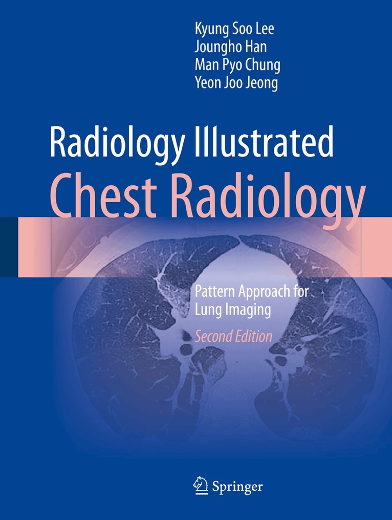
The purpose of this atlas is to illustrate how to achieve reliable diagnoses when confronted by the different abnormalities, or “disease patterns”, that may be visualized on CT scans of the chest. The task of pattern recognition has been greatly facilitated by the advent of high-technology CT such as helical and multidetector CT (MDCT) and dual-energy CT (DECT), and the focus of the book is very much on the role of state-of-the-art MDCT. A wide range of disease patterns and distributions are covered, with emphasis on the typical imaging characteristics of the various focal and diffuse lung diseases. In addition, clinical information relevant to differential diagnosis is provided and the underlying gross and microscopic pathology is depicted, permitting CT–pathology correlation. The entire information relevant to each disease pattern is also tabulated for ease of reference. This book will be an invaluable handy tool that will enable the reader to quickly and easily reach a diagnosis appropriate to the pattern of lung abnormality identified on CT scans. And in this second edition, some more signs and several new diseases and their pattern have been developed and updated. They will be provided with imaging, clinical relevance and CT-pathology correlation. Moreover, the chapters and disease patterns in their order of presentation shall be somewhat changed for readers’ easy legibility and understanding.
Häftad, Engelska, 2025
1 891 kr
Skickas inom 10-15 vardagar
The purpose of this atlas is to illustrate how to achieve reliable diagnoses when confronted by the different abnormalities, or “disease patterns”, that may be visualized on CT scans of the chest. The task of pattern recognition has been greatly facilitated by the advent of high-technology CT such as helical and multidetector CT (MDCT) and dual-energy CT (DECT), and the focus of the book is very much on the role of state-of-the-art MDCT. A wide range of disease patterns and distributions are covered, with emphasis on the typical imaging characteristics of the various focal and diffuse lung diseases. In addition, clinical information relevant to differential diagnosis is provided and the underlying gross and microscopic pathology is depicted, permitting CT–pathology correlation. The entire information relevant to each disease pattern is also tabulated for ease of reference. This book will be an invaluable handy tool that will enable the reader to quickly and easily reach a diagnosis appropriate to the pattern of lung abnormality identified on CT scans. And in this second edition, some more signs and several new diseases and their pattern have been developed and updated. They will be provided with imaging, clinical relevance and CT-pathology correlation. Moreover, the chapters and disease patterns in their order of presentation shall be somewhat changed for readers’ easy legibility and understanding.