
Anatomical Chart Company – författare
Visar alla böcker från författaren . Handla med fri frakt och snabb leverans.
21 produkter
21 produkter

404 kr
Skickas inom 7-10 vardagar
The Sixth Edition of Anatomy & Pathology: The World’s Best Anatomical Charts features 52 new and updated anatomical charts created by some of the world's best medical illustrators. This reference is an essential addition to every library, whether you are a health professional, student, or interested consumer. These anatomical charts show the human body in a format that provides a clear and visual understanding of human anatomy, physiology, and diseases. Medical terminology and easy-to-understand supporting text are printed directly on each chart so you never have to refer to a separate key card or manual. The convenient size and format make it ideal for studying, patient consultation or quick reference.Systems of the Body: 12Structures of the Body:16Diseases, Disorders and Conditions: 2458 pages52 full color chartssize 10" x 12"Spiral bound, soft cover

211 kr
Skickas inom 7-10 vardagar
The 3rd edition of Understanding Diabetes has been updated with new and additional information.Includes a general definition of diabetes.Describes how diabetes is diagnosed. Illustrates what happens in diabetes and insulin action in a body cell. Also describes the following types of diabetes:type I diabetestype 2 diabetes including factors causing insulin resistancepre-diabetesprenatal diabetesDiscusses diabetes in youth.Lists symptoms and risk factors. And includes tips for successful diabetes management.Available in the following size and formats:20" x 26" heavy paper ISBN 978-14698-9489-820" x 26" laminated with eyelets at top corners ISBN 978-14698-9492-8printed in USA

329 kr
Skickas inom 7-10 vardagar
The 3rd edition of Understanding Diabetes has been updated with new and additional information.Includes a general definition of diabetes.Describes how diabetes is diagnosed. Illustrates what happens in diabetes and insulin action in a body cell. Also describes the following types of diabetes:type I diabetestype 2 diabetes including factors causing insulin resistancepre-diabetesprenatal diabetesDiscusses diabetes in youth.Lists symptoms and risk factors. And includes tips for successful diabetes management.Available in the following size and formats:20" x 26" heavy paper ISBN 978-14698-9489-820" x 26" laminated with eyelets at top corners ISBN 978-14698-9492-8printed in USA

211 kr
Skickas inom 7-10 vardagar
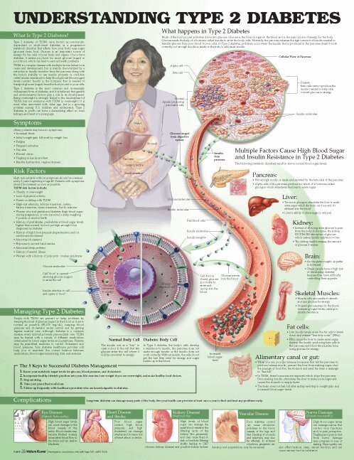
The third edition of our chart, Understanding Type 2 Diabetes uses patient friendly illustrations and text to explain type 2 diabetes and the complexity of type 2 diabetes. Defines type 2 diabetes and the role of glucose and insulin in the body Lists risk factors, symptoms and complications. Illustrates the many different factors that contribute to high blood sugar and insulin resistance. Also talks about management and gives management tips.Shows glucose molecules from the digestive system and insulin molecules from the pancreas traveling in a blood vessel. Illustrations visually compare a normal body cell to a body cell with diabetes and show when the cell develops a resistance to insulin, making it more difficult for glucose to enter the cell and leading to build-up of glucose in the blood vessel.The chart textually and visually presents complications from the disease: heart disease, stroke, vascular disease, nerve damage (neuropathy), kidney disease (nephropathy), periodontal disease (gum disease and mouth infection), and eye diseases such as glaucoma, cataracts, and diabetic retinopathy.Printed in USAsize 20" x 26"available in the following formats:heavy paper ISBN 978-1-4698-9497-3laminated with grommets at top corners ISBN 978-1-4698-9798-0
329 kr
Skickas inom 7-10 vardagar
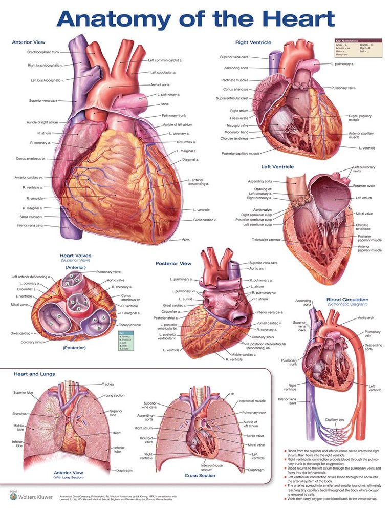
The Anatomy of the Heart anatomical chart, features full color, labeled illustrations clearly showing the anatomic structures and features of the heart.Illustrations include: Anterior View of the Heart Right Ventricle Left Ventricle Heart Valves Posterior View of the Heart Blood Circulation with explanation Anterior view of the Heart and Lungs Cross Section of the Heart and Lungs 20" x 26" heavy paper laminated with grommets at top corners
329 kr
Skickas inom 7-10 vardagar
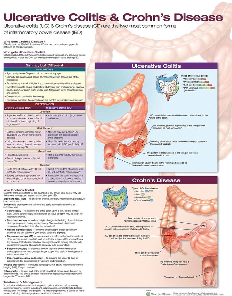
Ulcerative Colitis & Crohn's Disease

Häftad, Engelska, 2019
583 kr
Skickas inom 3-6 vardagar
Featuring more than 85 vibrant, fully annotated charts—19 new to this edition—this updated fourth edition of Diseases and Disorders: The World's Best Anatomical Charts is a perfect quick reference for medical and nursing students and an ideal visual aid for patient education. Printed on oversized, cardstock pages and compiled in a convenient, 10” x 12” spiral-bound volume with a laminated cover, these full-color charts created by some of the world's best medical illustrators illustrate and explain common diseases and disorders of the brain; heart; GI tract; eye and ear; endocrine, muscular, skeletal, reproductive, and respiratory systems; dental diseases; infectious diseases; healthy lifestyle issues; and cancer. Nineteen new charts cover Schizophrenia, Ovarian Cancer, Multiple Myeloma, Liver Cancer, Pancreatic Cancer, Prostate Cancer, Heart Failure, Peripheral Artery Disease, UC/Crohn's Disease, Irritable Bowel Syndrome, Age-Related Macular Degeneration, and more.All charts have been reviewed and updated.Medical terminology and easy-to-understand supporting text are printed directly on each chart.Every chart depicts an aspect of human anatomy, physiology, and disease presented in a clear, visual presentation.The book is ideal as a review resource or quick reference for studying human anatomy or for patient consultation and education.
329 kr
Skickas inom 7-10 vardagar
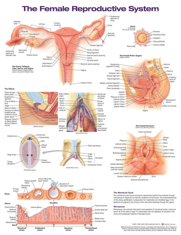
The Female Reproductive System anatomical chart features illustrated overviews of the perineum and pelvic organs; details of the ovary, fallopian tube, uterus, vagina, and clitoris; and changes to the ovary and uterus during phases of the menstrual cycle. The chart also describes the menstrual cycle and menopause. Illustrations included:The ovary, fallopian tube, uterus, and vagina (coronal section of aligned uterus, vagina, and proximal fallopian tube)Detail of an ovumThe female pelvic organs (sagittal section)The female perineum (inferior view, lithotomy position)NEW! The clitoris (median section and anteroinferior view)NEW! Detail of the clitoris (transverse section and anteroinferior view)The menstrual cycle (views of the ovary and uterus during each phase and ovulation)20" x 26" heavy paper laminated with grommets at top corners

Häftad, Engelska, 2022
1 093 kr
Skickas inom 7-10 vardagar
Travell, Simons & Simons’ Trigger Point Pain Patterns Flip Charts, Second Edition includes the iconic muscles and pain point patterns illustrations that set the standard in the field from Travell, Simons, & Simons' Myofascial Pain and Dysfunction: The Trigger Point Manual, the definitive reference on myofascial pain, and is organized in six sections following the structure of the Clinical Considerations chapters in the manual. This spiral-bound book with a built-in easel for display and patient presentation allows for a quick clinical reference to include TrPs as part of the clinical examination. Each section contains Trigger Point (TrP) pain referral patterns that may cause or be associated with a clinical condition commonly seen in clinical practice.
352 kr
Skickas inom 7-10 vardagar
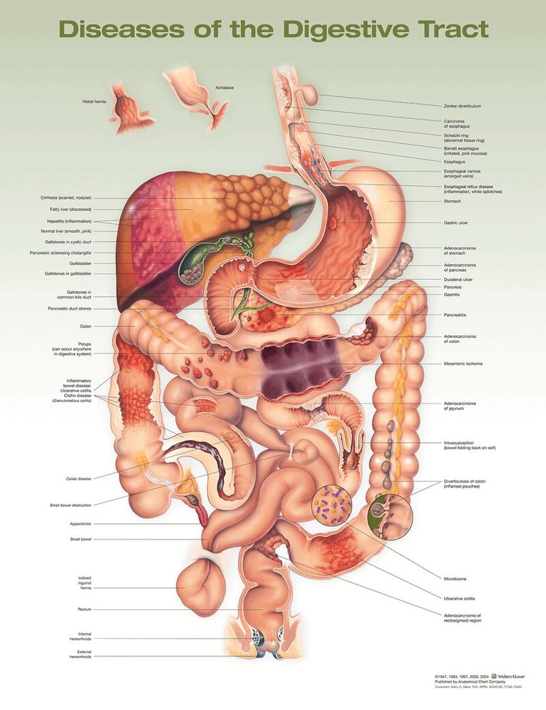
The updated Diseases of the Digestive Tract Anatomical Chart, 2nd Edition illustrates a wide variety of diseases using a central image of the organs of the digestive tract, part of the digestive system. Organs and approximately 40 diseases or conditions are clearly labeled, some with brief relevant descriptions, and illustrated in context. Included: Achalasia Adenocarcinoma of colon Adenocarcinoma of jejunum Adenocarcinoma of pancreas Adenocarcinoma of rectosigmoid region Adenocarcinoma of stomach Appendicitis Barrett’s esophagus Carcinoma of esophagus Celiac disease Cirrhosis Diverticulosis of colon Duodenal ulcer Esophageal reflux disease Esophageal varices External hemorrhoids Fatty liver Gallstones in common bile duct Gallstones in cystic duct Gallstones in gallbladder Gastric ulcer Gastritis Hepatitis Hiatal hernia Indirect inguinal hernia Inflammatory bowel disease: Ulcerative colitis, Crohn disease (Granulomatous Colitis)Internal hemorrhoids Intussusception Mesenteric ischemia Microbiome Pancreatic duct stones Pancreatic sclerosing cholangitis Pancreatitis Polyps Schatzki’s ring Small bowel obstruction Ulcerative colitis Zenker’s diverticulum 20" x 26" heavy paper laminated (without grommets), suitable for framing or hangingConsultant: Kathy A. Baker, PhD, APRN, ACNS-BC, FCNS, FAAN

336 kr
Skickas inom 7-10 vardagar
This update of the popular The Digestive System Anatomical Chart, 2nd Edition clearly illustrates the organs that make up the digestive system. All key structures are labeled, and the central image features the esophagus, liver, stomach (sectioned to show inside walls), gallbladder, pancreas, small and large intestines, rectum, appendix, arteries and veins. The chart also includes brief text sections describing the functions of the various organs in digestion. Additional, detailed illustrations include an orientation drawing of the digestive system in context of the human body along with: Muscles of Mastication Wall of Stomach Wall of Jejunum Wall of Colon Arterial Supply 20" x 26" heavy paper laminated (without grommets), suitable for framing or hangingOriginal medical illustrations by Brian Evan in consultation with Mark Frasier, Professor of Anatomy, Colorado State UniversityConsultant: Nicole R. Herring, PhD

227 kr
Skickas inom 7-10 vardagar
Ear, Nose, and Throat Anatomical Chart, 3rd Edition, is a highly illustrated chart providing clear, easy-to-understand information on the general anatomy of the region. Now with revised content, diversity of skin tones, and more detailed illustrations, it presents original artwork organized from ear, to nose, to throat, with each image clearly and fully labeled.Anatomical illustrations include: Large illustration highlighting the paranasal sinuses: frontal, ethmoid, and sphenoidal The ear with an inset of the tympanic membrane Oblique section of the cochlea, highlighting tympanic cavity, bony labyrinth, and membranous labyrinth Blood supply and innervation of the nasal cavity The mouth The tongue The larynx, lateral view Vocal cords, inspiration The pharynx, posterior view Swallowing, oral transit and pharyngeal phases
227 kr
Skickas inom 7-10 vardagar
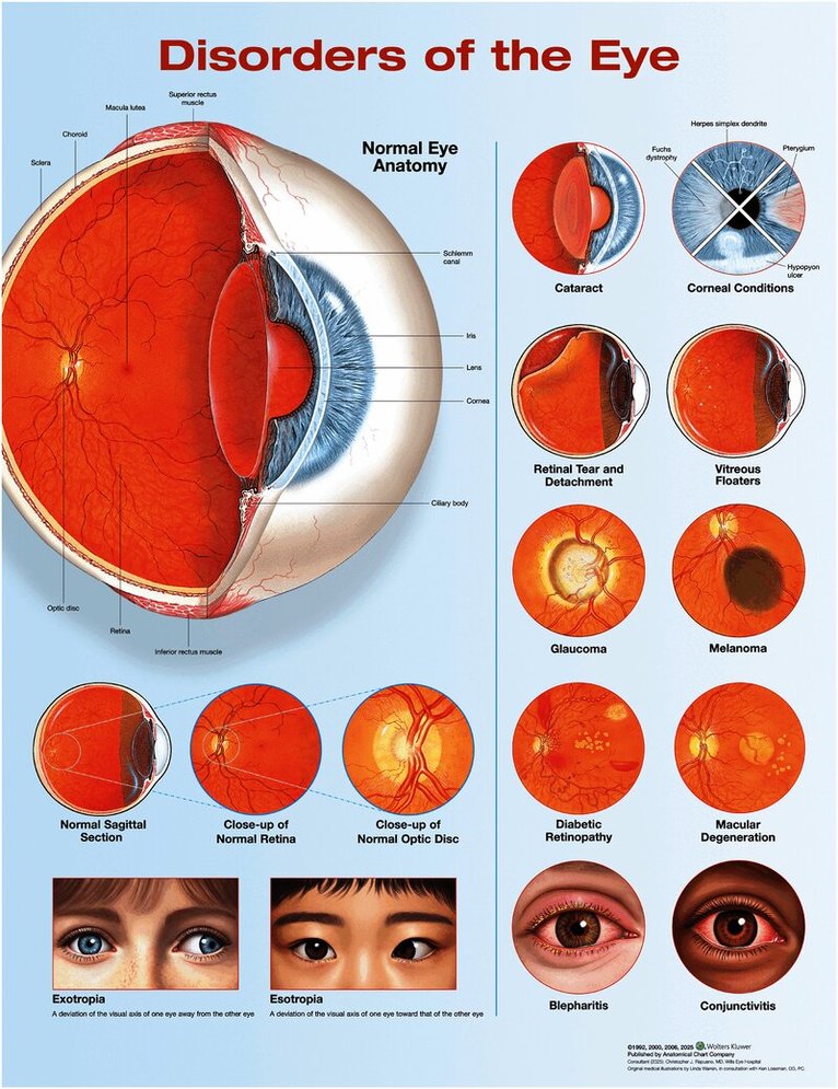
Disorders of the Eye Anatomical Chart, Third Edition features anatomy and clinical conditions of the eye with colorful detailed illustrations, all fully labeled, including diverse eye colors and skin tones. Normal eye anatomy, with cross section Cataract Corneal conditions Retinal tear and detachment Vitreous floaters Glaucoma Melanoma Diabetic retinopathy Macular degeneration Blepharitis and conjunctivitis Normal sagittal section with close-ups of normal retina and optic disc Exotropia and esotropia 20" x 26" heavy paper laminatedConsultant (2025): Christopher J. Rapuano, MD, Wills Eye HospitalOriginal medical illustrations by Linda Warren, in consultation with Ken Lossman, PC
227 kr
Skickas inom 7-10 vardagar
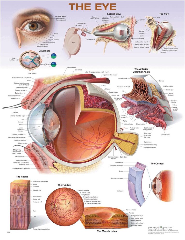
The Eye Anatomical Chart, Second Edition features general anatomy of the eye with colorful detailed illustrations, all fully labeled. Outer eye with surface anatomy, anterior view, also showing the lacrimal gland and nasolacrimal duct Eyeball in skull, lateral and top views Visual field diagram Cross section of the eye, lateral view, as the large central illustration along with the anterior chamber angle Medial cross section of the eye The cornea, macula lutea, fundus, and retina in close-up views 20" x 26" heavy paper laminatedConsultant (2025): Christopher J. Rapuano, MD, Wills Eye HospitalOriginal medical illustrations by Keith Kasnot, CMI, in consultation with Randall Paul, OD, Phoenix, Arizona
226 kr
Tillfälligt slut
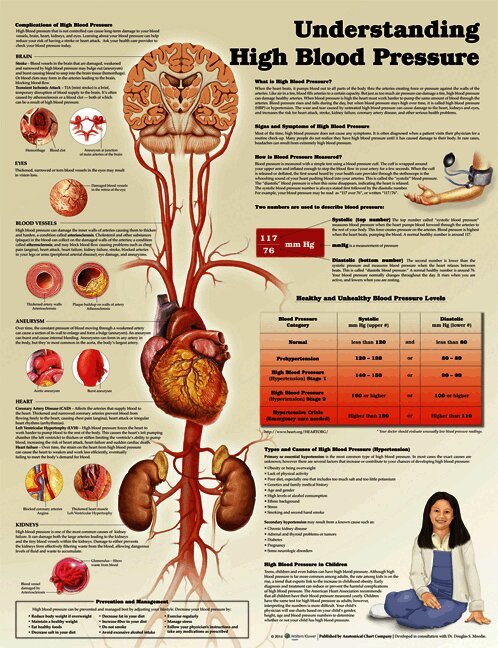
Understanding High Blood Pressure explains and illustrates the how and why of HBP with clear easy to understand text, tables and imagery. This chart explains what high blood pressure is, how its measured, healthy and unhealthy blood pressure levels, types and caused of high blood pressure, symptoms of high blood pressure, complications and prevention and management tips. Also discusses high blood pressure in childrenmade in USAsize 20" x 26" Available in the following versions: heavy paper ISBN 9781469872889laminated with grommets at the top for hanging ISBN 9781469872896
358 kr
Tillfälligt slut
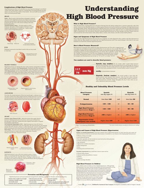
Understanding High Blood Pressure explains and illustrates the how and why of HBP with clear easy to understand text, tables and imagery. This chart explains what high blood pressure is, how its measured, healthy and unhealthy blood pressure levels, types and caused of high blood pressure, symptoms of high blood pressure, complications and prevention and management tips. Also discusses high blood pressure in children.made in USAsize 20" x 26" Laminated with grommets at top for hangingAvailable in the following versions: heavy paper ISBN 9781469872889laminated with grommets at the top for hanging ISBN 9781469872896

233 kr
Tillfälligt slut
The Third Edition of our chart Understanding Type 1 Diabetes has been updated with additional content and new illustrations. It provides easy-to-understand visuals and text descriptions of how Type 1 diabetes affects the process of insulin production by the pancreas, which in turn affects glucose conversion to energy. Included is a side-by-side comparison of a normal body cell and a diabetic body cell, illustrating how they differ in converting glucose from food to energy.The chart describes the importance of controlling the level of glucose in the blood and provides tips for management of type 1. It lists symptoms of short-term complications such as hypoglycemia, hyperglycemia, and ketoacidosis. It also describes long-term complications of the disease.Size 20" x 26"printed in USAAvailable in the following versions:heavy paper ISBN 978-1-4698-9493-5laminated with eyelets at top corners ISBN 978-1-4698-9494-2

358 kr
Tillfälligt slut
The Third Edition of our chart Understanding Type 1 Diabetes has been updated with additional content and new illustrations. It provides easy-to-understand visuals and text descriptions of how Type 1 diabetes affects the process of insulin production by the pancreas, which in turn affects glucose conversion to energy. Included is a side-by-side comparison of a normal body cell and a diabetic body cell, illustrating how they differ in converting glucose from food to energy.The chart describes the importance of controlling the level of glucose in the blood and provides tips for management of type 1. It lists symptoms of short-term complications such as hypoglycemia, hyperglycemia, and ketoacidosis. It also describes long-term complications of the disease.Size 20" x 26"printed in USAAvailable in the following versions:heavy paper ISBN 978-1-4698-9493-5laminated with eyelets at top corners ISBN 978-1-4698-9494-2
358 kr
Tillfälligt slut
The third edition of our chart, Understanding Type 2 Diabetes uses patient friendly illustrations and text to explain type 2 diabetes and the complexity of type 2 diabetes. Defines type 2 diabetes and the role of glucose and insulin in the body Lists risk factors, symptoms and complications. Illustrates the many different factors that contribute to high blood sugar and insulin resistance. Also talks about management and gives management tips.Shows glucose molecules from the digestive system and insulin molecules from the pancreas traveling in a blood vessel. Illustrations visually compare a normal body cell to a body cell with diabetes and show when the cell develops a resistance to insulin, making it more difficult for glucose to enter the cell and leading to build-up of glucose in the blood vessel.The chart textually and visually presents complications from the disease: heart disease, stroke, vascular disease, nerve damage (neuropathy), kidney disease (nephropathy), periodontal disease (gum disease and mouth infection), and eye diseases such as glaucoma, cataracts, and diabetic retinopathy.Printed in USAsize 20" x 26"available in the following formats:heavy paper ISBN 978-1-4698-9497-3laminated with grommets at top corners ISBN 978-1-4698-9798-0
358 kr
Tillfälligt slut
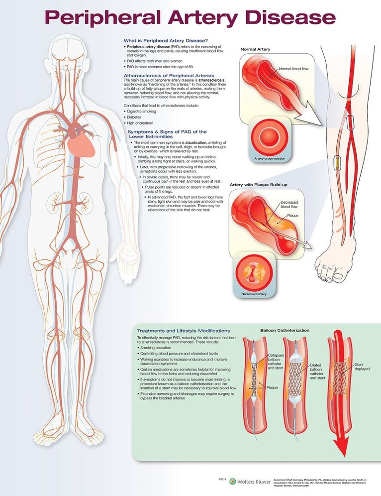
Laminated Chart of the Peripheral Artery Disease

Häftad, Engelska, 2002
289 kr
Tillfälligt slut