
Auguste Wackenheim – författare
Visar alla böcker från författaren . Handla med fri frakt och snabb leverans.
13 produkter
13 produkter
Häftad, Engelska, 1982
1 116 kr
Skickas inom 10-15 vardagar
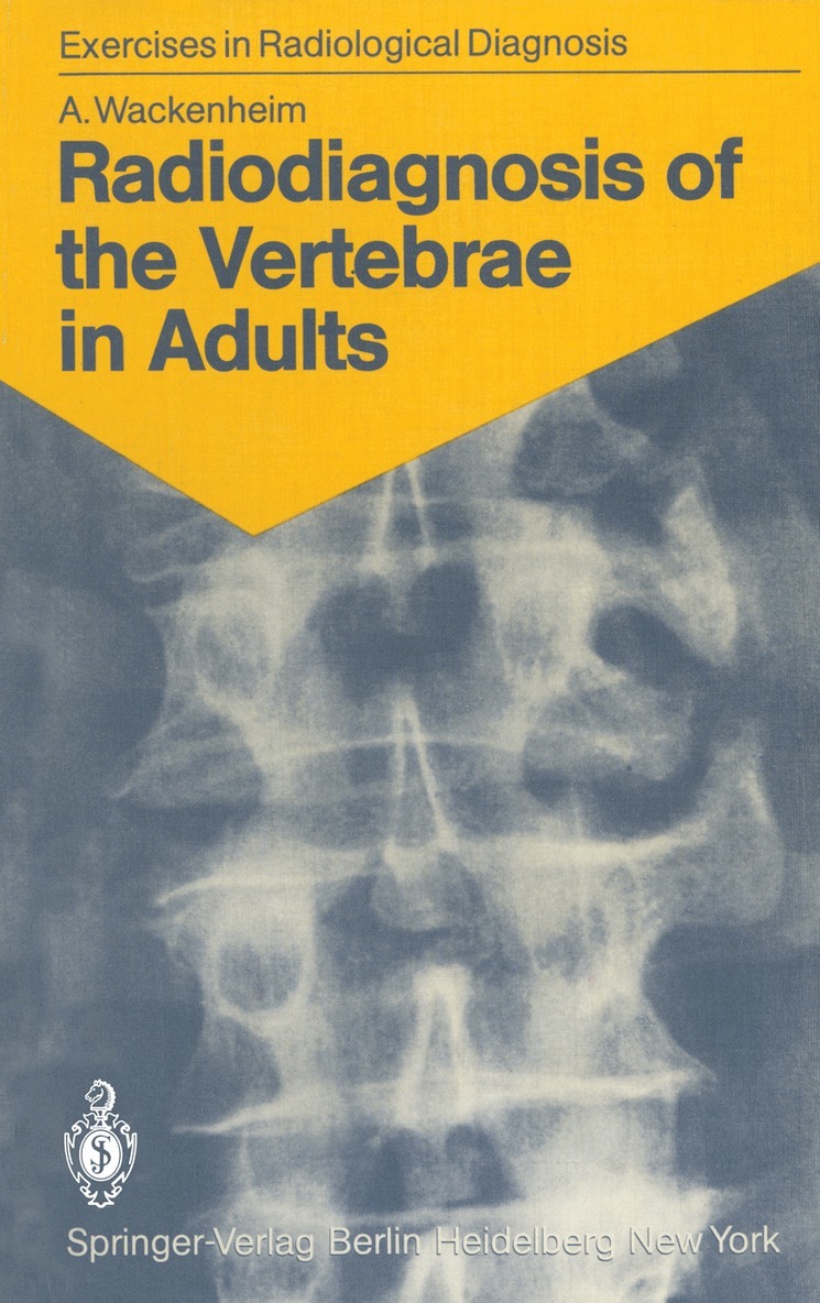
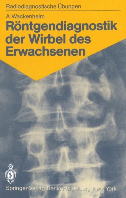
This book is intended for beginners and for those who want to refresh their knowledge of the elementary radioanatomy of the vertebrae, particularly their pathological radioanatomy. I do not pretend, as does Roger Martin du Gard's hero, that one always must begin with a radiographic examination, but I do believe that a student, especially one interested in radiology, must be able to apprehend an image isolated from its clinical context. To optimize memorization of the image I have selected unmistakable cases with marked, well-evolved lesions. This will enable the studentlater to recognize less distinct images of the same kind. The first section of the book is exclusively iconographic. After studying an image, the reader will find in the second section, under the appropriate reference number, a commen tary illustrated with a realistic drawing by my friend Dr. Csaba Hethalmi. Attention to the following points will assist a fruitful reading: 1. Cases 1-5 involve normal subjects; all other cases are pathological. 2. The reader must imagine that he is conducting a routine examination and draw on his resources to make a practical analysis of an image. As a matter of fact, all the films (except that in case 46, which is the radiograph of a specimen) were -indeed taken under routine conditions using standard pro jections. 3. The cases are in no systematic or nosological order. Each case is illustrated with one, two, or (rarely) three images.
Häftad, Tyska, 1982
577 kr
Skickas inom 10-15 vardagar
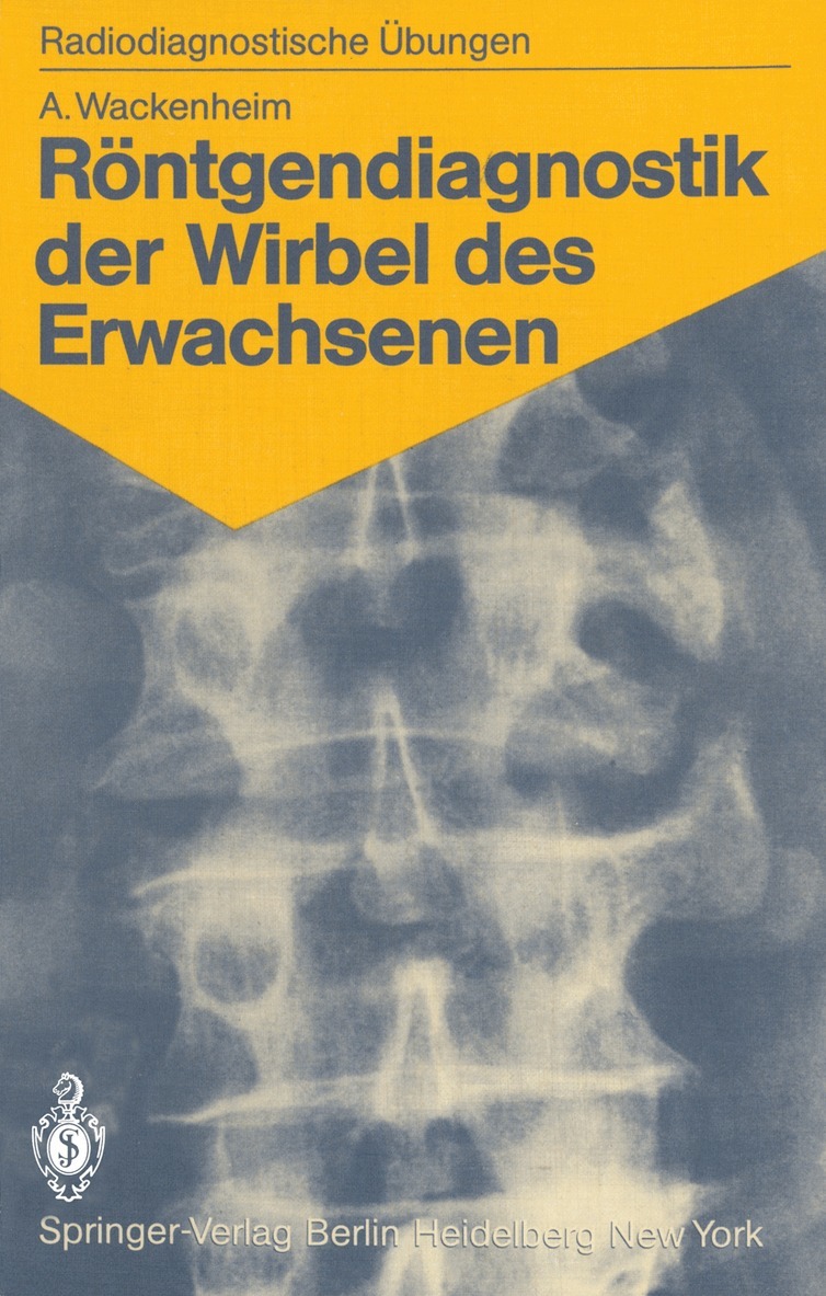
Der Schwerpunkt dieses Buches liegt auf dem Erkennen und der Interpretation r|ntgenologischer Zeichen. Der Leser soll da- durch in die Lage versetzt werden, die Bildsprache einer R|ntgenaufnahme zu erkennen. Die r|ntgenologischen Zeichen werden jeweils in einem dreistufigen Informationsgang vorge- stellt. Die Autoren stellen jeweils die charakteristischen, die spezifischen und die pathognomonischen Zeichen vor und geben ihre diagnostische Wertigkeit an.
Häftad, Engelska, 1985
1 116 kr
Skickas inom 10-15 vardagar
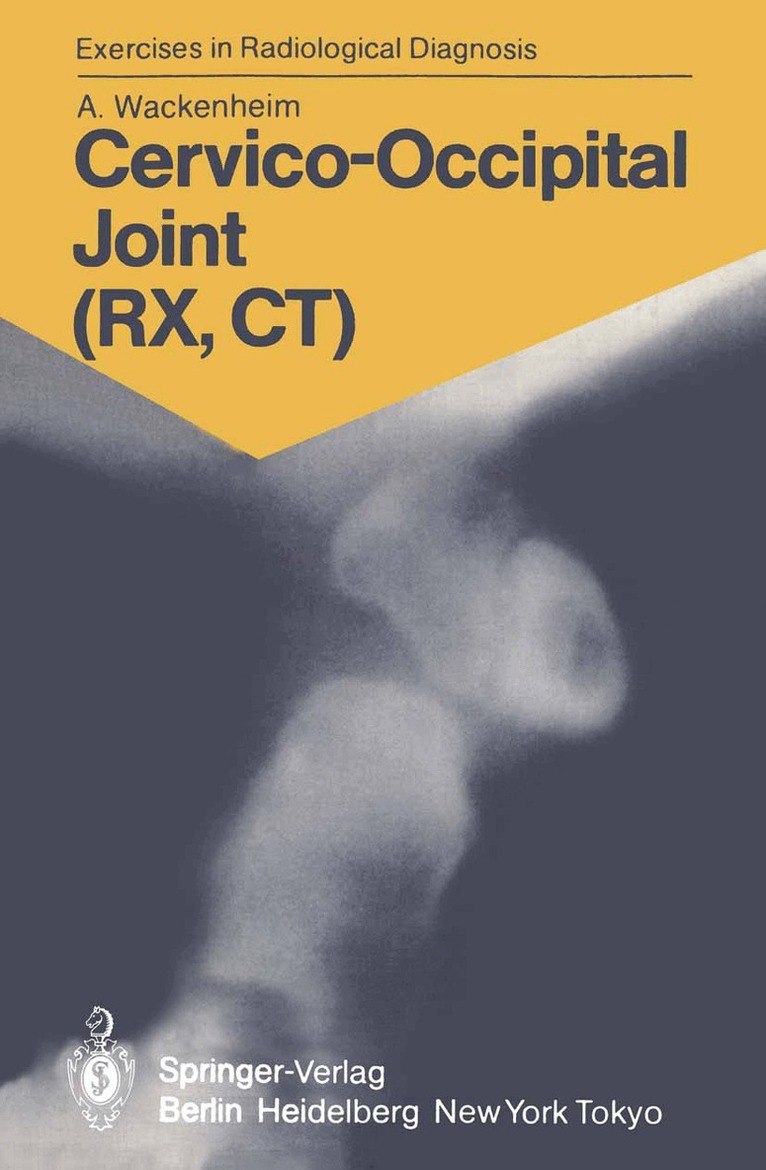
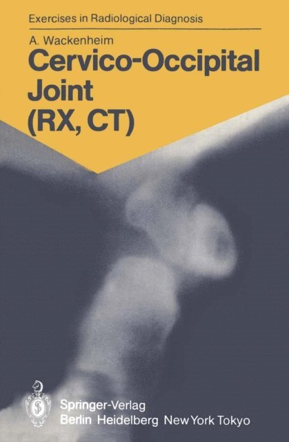
Like my first book of exercises in radiological diagnosis con- cerning the vertebrae in adults, the first part of this book consists of an atlas of images ready for immanent reading. The images are numbered, each number corresponding to a case. The second part comprises the corresponding comments, critical interpretations, and drawings (using the same num- bers as the corresponding radiographs in the first part). I hope that this second booklet will be as appreciated as was the first, which was published in four languages (Radio- diagnosis of the Vertebrae in Adults. Springer, Berlin Hei- delberg New York 1983). My own documentation having proved deficient in some fields, I had recourse to the didactic collections oimy collea- gues, friends, and students. Special thanks are due to Prof. J. F. Bonneville, and to Drs. J. L. Dietemann, Y. Dirheimer, J. C. Dosch, and J. Vignaud. AUGUSTE WACKENHEIM VII Contents Introduction ...1 Part One: Iconography 5 Part Two: Commentary with Corresponding Schemata. 107 References . .189 Subject Index 191 IX Introduction The analysis or the reading of an X-ray picture proceeds from struc- turalistic rules, such as the immanence, the synchronism, the significa- tion (signifiant and signifie of a sign), first from the semeiologic and then from the semantic point of view. In a simpler way, one can say that it is necessary to distinguish the signifiant, the signiGBPie, the comment, the interpretation, and the radiobioclinical confrontation. The signifiant or character is a normal or pathological "characteris- tic" part of the image.
Häftad, Tyska, 1985
577 kr
Skickas inom 10-15 vardagar
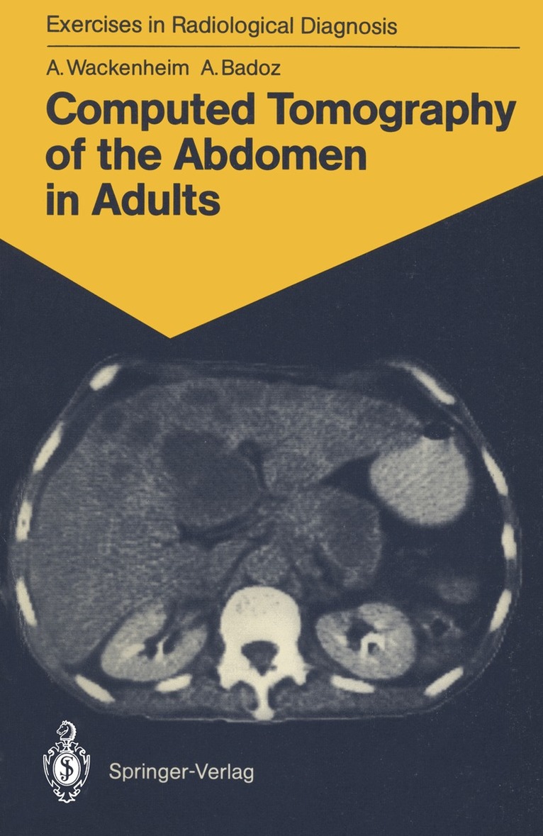
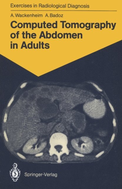
Computed Tomography of the Abdomen in Adults
85 Radiological Exercises for Students and Practitioners
Häftad, Engelska, 1988
561 kr
Skickas inom 10-15 vardagar
These exercises are meant for students and practitioners who wish to familiarize themselves with the normal and pathologieal computerized tomographie radioanatomy of the abdomen. The iconography is suffieiently characteristic to be read without the help of clinical or biological data. It comprises both normal and pathologie findings. Analysis of scans is comprised of two steps. The first part consists of the detailed study of normal scans, whieh serve as a reference. For this, eight main slice levels have been considered necessary and sufficient: neces sary since a certain number of slices are indispensable for the exploration of the abdomen; sufficient because a larger number of slices would risk rendering memorization difficult. The second part involves a study of the pathologie findings, organ by organ. Acknowledgements. Appreciation is extended to all those who have helped in realizing this study and, more particularly, to our friends and colleagues, J. L. DIETEMANN, C. Roy, J. L. BURGUET, M. VOUGE, and J. W. SOUITER. We would also like to thank Dr. J. WIECZOREK for his friendly assistance and advice in the planning and presentation of figures and schemata. 1 Technical Note Computerized tomography of the abdomen begins with an initial image called "scout view". This numbered radio graph of the abdomen is an analogous representation of the information and allows the location of the eight selected slice levels; these are represented by horizontallines. The slices are 10 mm thick and are taken at intervals of 2.5 cm.
Häftad, Tyska, 1988
577 kr
Skickas inom 10-15 vardagar
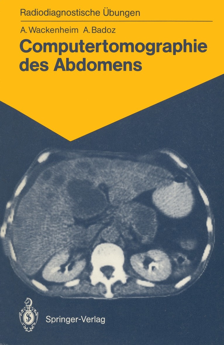
Dieses Übungsbuch richtet sich an Studenten, Ärzte und Radiologen, die sich mit der Auswertung von Computertomogrammen des Abdomens vertraut machen wollen. Die einzelnen Fälle zeigen zum einen Kontrollaufnahmen des Gesunden und zum anderen pathologische Veränderungen, die organspezifisch klassifiziert sind. Der Leser wird im vorliegenden Band schrittweise an die systematische Interpretation der Computertomogramme herangeführt.

E-bok
PDF, Engelska, 20121 459 kr
Läs direkt efter köp
This book is intended for beginners and for those who want to refresh their knowledge of the elementary radioanatomy of the vertebrae, particularly their pathological radioanatomy. I do not pretend, as does Roger Martin du Gard''s hero, that one always must begin with a radiographic examination, but I do believe that a student, especially one interested in radiology, must be able to apprehend an image isolated from its clinical context. To optimize memorization of the image I have selected unmistakable cases with marked, well-evolved lesions. This will enable the studentlater to recognize less distinct images of the same kind. The first section of the book is exclusively iconographic. After studying an image, the reader will find in the second section, under the appropriate reference number, a commen tary illustrated with a realistic drawing by my friend Dr. Csaba Hethalmi. Attention to the following points will assist a fruitful reading: 1. Cases 1-5 involve normal subjects; all other cases are pathological. 2. The reader must imagine that he is conducting a routine examination and draw on his resources to make a practical analysis of an image. As a matter of fact, all the films (except that in case 46, which is the radiograph of a specimen) were -indeed taken under routine conditions using standard pro jections. 3. The cases are in no systematic or nosological order. Each case is illustrated with one, two, or (rarely) three images.
E-bok
PDF, Tyska, 2013582 kr
Läs direkt efter köp
E-bok
PDF, Engelska, 20121 459 kr
Läs direkt efter köp
E-bok
PDF, Tyska, 2013582 kr
Läs direkt efter köp
Computed Tomography of the Abdomen in Adults
85 Radiological Exercises for Students and Practitioners
E-bok
PDF, Engelska, 2012708 kr
Läs direkt efter köp
These exercises are meant for students and practitioners who wish to familiarize themselves with the normal and pathologieal computerized tomographie radioanatomy of the abdomen. The iconography is suffieiently characteristic to be read without the help of clinical or biological data. It comprises both normal and pathologie findings. Analysis of scans is comprised of two steps. The first part consists of the detailed study of normal scans, whieh serve as a reference. For this, eight main slice levels have been considered necessary and sufficient: neces sary since a certain number of slices are indispensable for the exploration of the abdomen; sufficient because a larger number of slices would risk rendering memorization difficult. The second part involves a study of the pathologie findings, organ by organ. Acknowledgements. Appreciation is extended to all those who have helped in realizing this study and, more particularly, to our friends and colleagues, J. L. DIETEMANN, C. Roy, J. L. BURGUET, M. VOUGE, and J. W. SOUITER. We would also like to thank Dr. J. WIECZOREK for his friendly assistance and advice in the planning and presentation of figures and schemata. 1 Technical Note Computerized tomography of the abdomen begins with an initial image called "scout view". This numbered radio graph of the abdomen is an analogous representation of the information and allows the location of the eight selected slice levels; these are represented by horizontallines. The slices are 10 mm thick and are taken at intervals of 2.5 cm.
Häftad, Tyska, 1970
577 kr
Skickas inom 10-15 vardagar
E-bok
PDF, Tyska, 2013566 kr
Läs direkt efter köp