
Brian Heaton – författare
Visar alla böcker från författaren Brian Heaton. Handla med fri frakt och snabb leverans.
3 produkter
3 produkter
Häftad, Engelska, 2017
1 050 kr
Skickas inom 10-15 vardagar
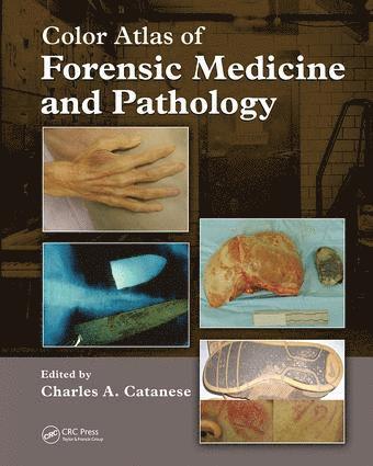
New York City has the largest medical examiner’s office in the United States, and the Brooklyn division is the busiest of the five boroughs. Charles A. Catanese received his Forensic Pathology fellowship training in New York, and then worked full time as a Medical Examiner in the Brooklyn office for more than 10 years. He has personally performed more than 4000 autopsies, including over 400 homicides. Dr. Catanese has worked through several disasters, including TWA Flight 800, AA Flight 587, and more than nine months on the World Trade Center fatalities. He is currently the Chief Medical Examiner of Orange County, New York. Drawing on his wealth of knowledge and experience in solving some of the most difficult cases a forensic examiner could encounter, he assembles hundreds of images from his own work experience to present the Color Atlas of Forensic Medicine and Pathology. Featuring twice the number of images as any other forensic pathology atlas, the book is filled with high-resolution photos that demonstrate postmortem changes of the human body and the different types of patterns produced in deaths caused by: Natural causesDiagnostic or therapeutic proceduresSubstance abuse PoisoningChild abuseFirearmsBlunt instrumentsSharp instrumentsBurnsAsphyxiaThis easy-to-read atlas, created for medical and non-medical personnel, covers basic and advanced forensic concepts that relate to all manners of deaths. The carefully worded, unambiguous text describing each photo and the side-by-side comparisons of similar, yet different, pathologies make this remarkable atlas a powerful teaching tool for all those who must confront and solve the mystery of human demise. A fully searchable DVD version is also available.
Inbunden, Engelska, 2011
1 351 kr
Skickas inom 10-15 vardagar
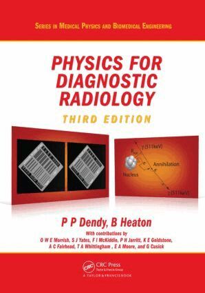
With every chapter revised and updated, Physics for Diagnostic Radiology, Third Edition continues to emphasise the importance of physics education as a critical component of radiology training. This bestselling text helps readers understand how various imaging techniques work, from planar analogue and digital radiology to computed tomography (CT), nuclear medicine, and positron emission tomography (PET) to ultrasound imaging and magnetic resonance imaging (MRI).New to the Third Edition Material on digital receptorsEmphasis on the differences between analogue and digital imagesCoverage of multi-slice CT and three-dimensional resolution, dual energy applications, and cone beam CTSpecial radiographic techniques, including subtraction techniques and interventional radiologyNew chapter on PET, with discussion of multi-modality imaging (PET/CT)Additional material on radiation doses and risks to patientsNew chapter covering picture archiving and communication system (PACS), teleradiology, networks, archiving, and related factorsA summary of the main teaching points at the beginning of each chapterAfter an introductory chapter on basic physics, the book follows the x-ray imaging process: production of x-rays, interaction with the patient, radiation measurement, the image receptor, the radiological image, and image quality assessment. It then covers more advanced x-ray techniques as well as imaging with radioactive materials. The text also focuses on radiobiology, risk and radiation protection, and imaging with non-ionising radiation. The final chapter discusses data handling in a modern, electronic radiology department.
Häftad, Engelska, 2005
2 415 kr
Tillfälligt slut