
Carlo Bartolozzi – författare
2 223 kr
Skickas inom 10-15 vardagar
2 925 kr
Läs direkt efter köp
Few fields of medicine have witnessed such impressive progress as the diagnosis and treatment of liver tumors. Advances in imaging technology, the development of novel contrast agents, and the introduction of optimized scanning protocols have greatly facilitated the non-invasive detection and characterization of focal liver lesions. Furthermore, image-guided techniques for percutaneous tumor ablation have become an accepted alternative treatment for patients with inoperable liver cancer. This book provides a comprehensive and up-to-date overview of the role of diagnostic and interventional radiology in respect of liver tumors. The volume moves from background sections on methodology and segmental liver anatomy to the main sections on the diagnosis of benign and malignant liver lesions. An integrated approach, focused on the correlation of ultrasound, CT, and MR imaging findings, is presented. Finally, a full section describes the principles, methods, and results of percutaneous tumor ablation techniques.
2 925 kr
Läs direkt efter köp
Few fields have witnessed such impressive advances as image processing in radiology. The progress achieved has revolutionized diagnosis and greatly facilitated treatment selection and accurate planning of procedures. This book, written by leading experts from many countries, provides a comprehensive and up-to-date description of how to use 2D and 3D processing tools in clinical radiology. The first section covers a wide range of technical aspects in an informative way. This is followed by the main section, in which the principal clinical applications are described and discussed in depth. To complete the picture, a third section focuses on various special topics. The book will be invaluable to radiologists of any subspecialty who work with CT and MRI and would like to exploit the advantages of image processing techniques. It also addresses the needs of radiographers who cooperate with clinical radiologists and should improve their ability to generate the appropriate 2D and 3D processing.
2 223 kr
Skickas inom 10-15 vardagar

2 223 kr
Skickas inom 10-15 vardagar

2 777 kr
Skickas inom 10-15 vardagar
1 115 kr
Skickas inom 10-15 vardagar
1 459 kr
Läs direkt efter köp
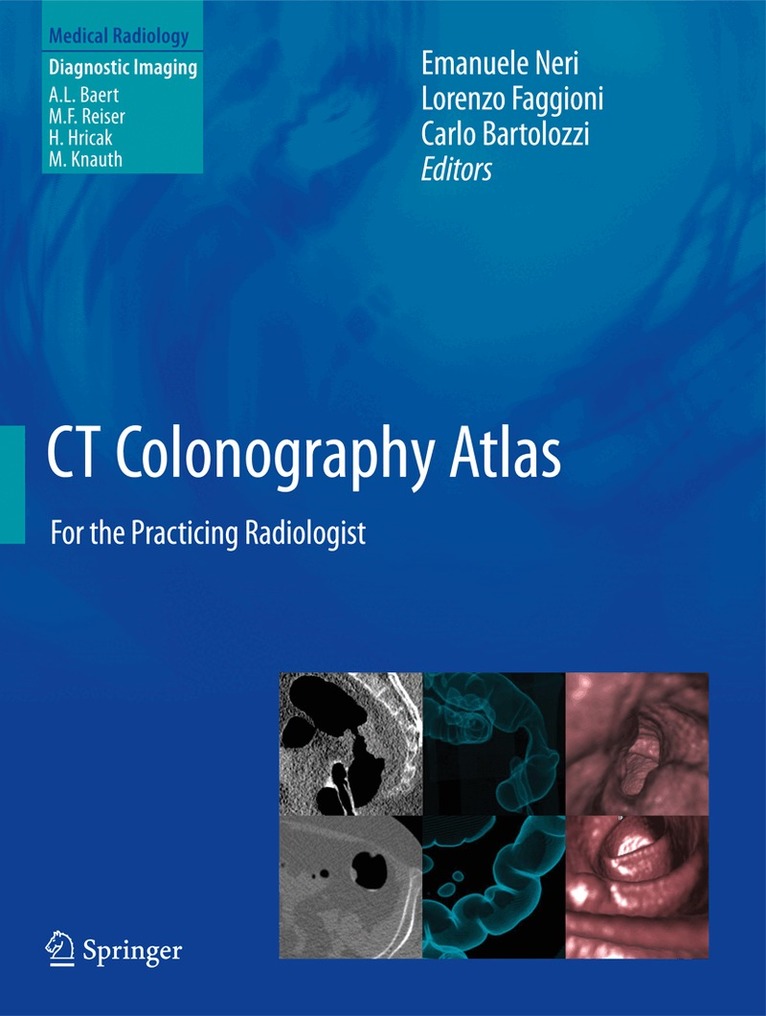
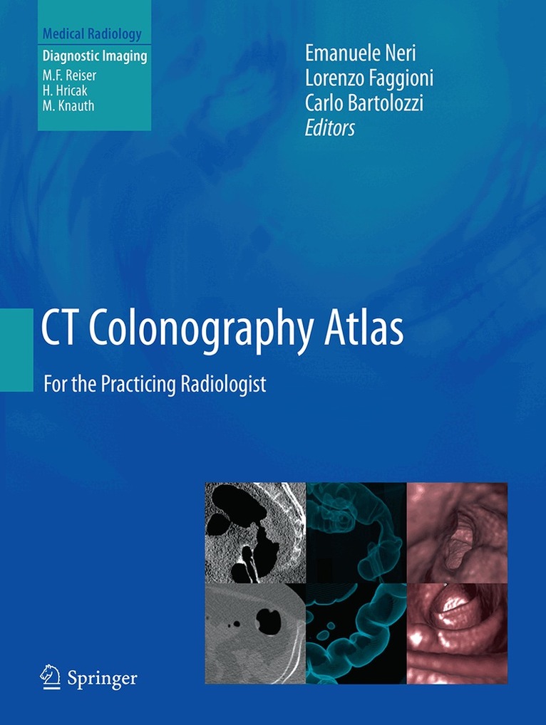
This easy-to-use atlas comprises a collection of representative common and unusual virtual colonoscopy (CT colonography, CTC) cases that physicians and radiologists may expect to encounter during their clinical practice. The atlas reflects the important recent advances in image acquisition, patient preparation, and image processing and is thus completely up-to-date. Each case is presented with the native CT images, integrated images obtained by 3D image processing, and colonoscopic correlation. Topics covered include normal appearances, anatomical variants, pitfalls, diverticula, lipomas, inflammatory bowel disease, polyps, flat lesions, cancers, and the postsurgical colon. By presenting the main features of anatomy and pathology, this atlas will serve as an invaluable tool both for radiologists performing CTC and for clinicians who need to review the CTC examinations of their patients.
2 925 kr
Läs direkt efter köp
This book provides a lucid summary of modern multislice CT imaging of the abdomen, with a focus on the essential imaging findings. After a concise technical introduction, the most important abdominal diseases are described and illustrated with high-quality images. Sections are devoted to the liver and biliary system, the pancreas and spleen, the kidneys and urogenital system, and the bowel and peritoneal cavity. Throughout, key differential diagnostic features are highlighted. The editorial team is composed of internationally renowned radiologists from Europe and the United States, and all chapters have been written by recognized experts in the topic under consideration. Multislice CT of the Abdomen will serve as an excellent reference for radiologists participating in further professional training and will prove an ideal source of information for all who wish to deepen their personal knowledge of the subject.
2 223 kr
Skickas inom 10-15 vardagar
1 336 kr
Skickas inom 10-15 vardagar
1 115 kr
Skickas inom 10-15 vardagar
1 459 kr
Läs direkt efter köp
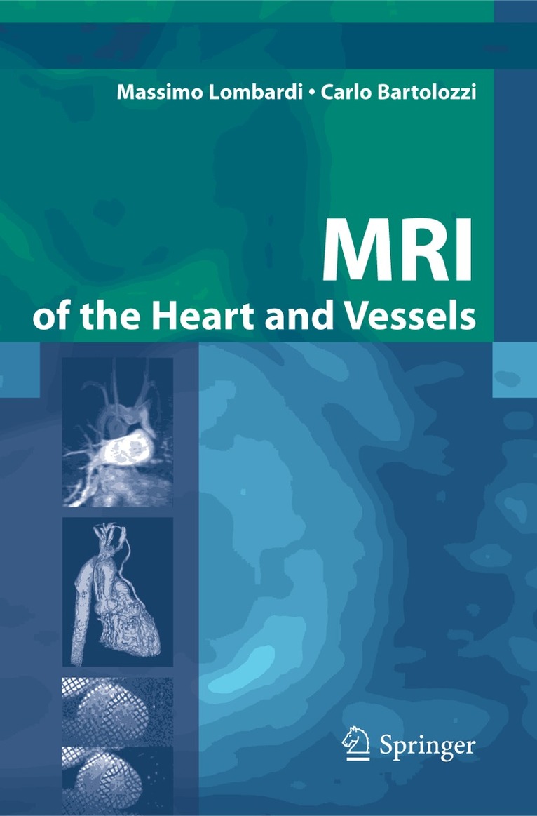
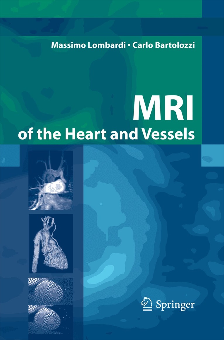
In recent years magnetic resonance imaging (MRI) has enriched the technological potential available for the characterization of cardiovascular pathologies, adding substantial advantages to other non-invasive techniques. This technique, which is intrinsically digital and has reduced operator dependency, allows the performance of image analysis in a quantitative and reproducible manner.
The use of non-ionizing energy with the consequent absence of an environmental impact and of operator and patient biohazards makes MRI a winning technique when evaluating the risk – benefit ratio in comparison to other imaging methods.
In virtue of its added diagnostic value and inherent refinements that allow construction of two- and three-dimensional images, MRI is gaining a primary role in the histopathological and physiopathological understanding of a large number of pathologies concerning the heart and vessels.
This text is addressed both to MRI operators seeking specific technical information and to clinicians who wish to have a better understanding of the diagnostic and management advantages that MRI can offer.
1 669 kr
Skickas inom 10-15 vardagar