
Carol B. Benson – författare
206 kr
Läs direkt efter köp
Awarded the iParenting Media Award for Excellent Product of 2008!
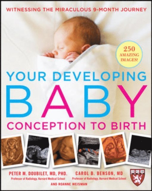
Experience the miracle of life-with your own eyes
Congratulations, parents-to-be! You''re about to embark on a momentous journey. Even more exciting, you''re the first generation of parents who-thanks to 3D and conventional ultrasound-can actually “see” your child before he or she is born. This wonderful, one-of-a-kind guide, written by two Harvard professors, takes you through every stage of your baby''s development, from conception to delivery-with more than 200 images and drawings to illustrate each glorious moment.
Get a guided tour of a baby''s journey with:
Breathtaking 3D images of babies'' faces, limbs, and other featuresDetailed 2D ultrasounds of developing organs and bones inside the bodyVisual pointers on what to look for, and what you''re seeing, on the screenWith this illuminating guide, you''ll be able to see how doctors determine if your baby is a boy or girl and whether you''re having twins or multiples. You''ll be able to watch the growth of your baby''s hands, feet, heart, head, and more. Best of all, you''ll be able to actively participate in the greatest journey of all: from single cell to bringing a new baby into the world.
Visit www.YourDevelopingBaby.com for more information.
Library Journal starred review: "Using 250 diagnostic ultrasound images, Harvard radiology professors Doubilet and Carol B. Benson present a marvelous book charting the growth of babies in the womb. . . . The authors well explain the different types of ultrasound and their medical uses, especially the 3D images that show the baby’s outer surface and the 2D images revealing the internal development of organs. This virtual tour of a life in the making will attract future parents in droves."--Janet M. Schneider, James A. Haley Veterans’ Hospital, Tampa, FL
Sciencenews.org:" . . . an accessible and captivating text that guides readers through 9 months of pregnancy."
3 078 kr
Skickas inom 3-6 vardagar
797 kr
Läs direkt efter köp
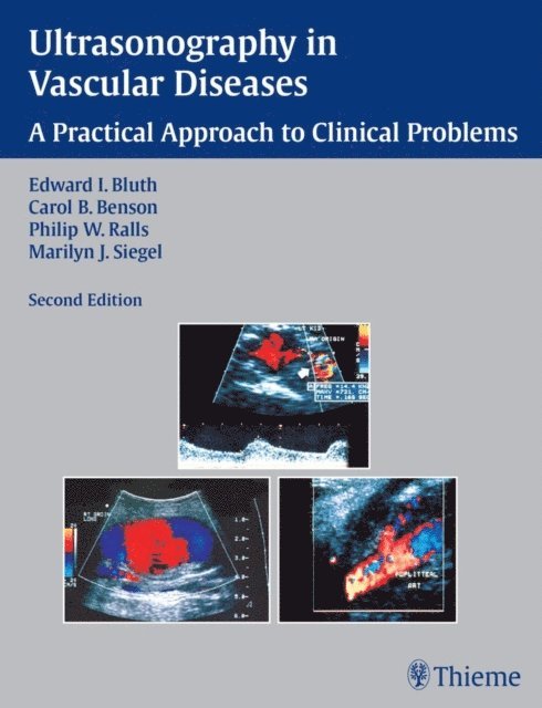
An invaluable resource for ultrasound imaging for vascular diseases
Ultrasonography in Vascular Diseases: A Practical Approach to Clinical Problems is a concise guide to the latest clinical applications of ultrasound in diagnosing vascular disorders and diseases. Well-known authorities in the field provide straightforward instruction on how to choose the appropriate imaging examination and complete the imaging workup of the patient for the full range of vascular problems.
Highlights:
Practical information on the usefulness of ultrasound, non-imaging tests, or other imaging modalities, such as CT and MR Thorough descriptions of symptoms, differential diagnosis, techniques, as well as the possible complications, benefits, and limitations of each technique More than 150 images and photographs illustrate key conceptsIdeal for reference and review, this text will prove to be an indispensable clinical reference for ultrasonographers, radiologists, interventional radiologists, vascular surgeons, cardiologists, vascular medicine specialists, residents, physicians, nurses, and radiology assistants.
2 684 kr
Läs direkt efter köp
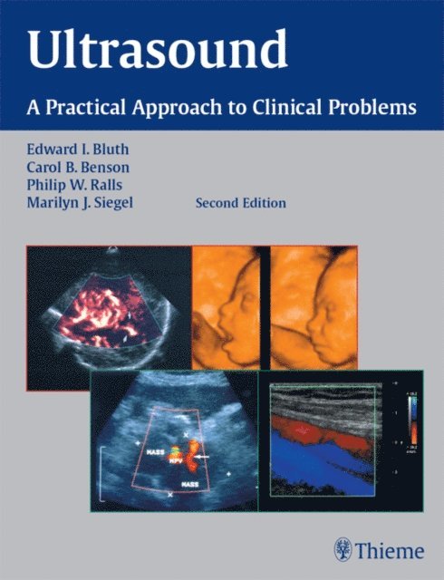
A practical reference for completing the imaging workup of the patient
Based on a popular course taught at the Radiological Society of North America''s Annual Meeting, this book provides all the essential information for choosing the appropriate imaging examination and completing the imaging workup of a patient. Chapters are organized into parts according to the anatomical location of the clinical problems addressed. The authors guide the reader through the diagnostic evaluation, reviewing the indications for and the strengths and limitations of ultrasound imaging.
Features:
Practical information on the usefulness of ultrasound, non-imaging tests, or other imaging modalities, such as CT and MR, for evaluating each clinical situation Clear descriptions of symptoms and differential diagnosis Nearly 1,300 images and photographs demonstrating key points A new chapter on neonatal spinal cord anomaliesComprehensive and up-to-date, this edition is essential for ultrasonographers, radiologists, residents, physicians, nurses, and radiology assistants seeking the latest recommendations for the effective use of ultrasonography.
1 108 kr
Läs direkt efter köp
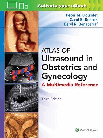
The latest recommendations for using ultrasound in obstetrics and gynecology
From diagnosing pelvic pain and bleeding, to the use of ultrasound in screening and treatments for ovarian cancer, infertility, and maternal complications of diabetes mellitus, this book covers the full spectrum of clinical applications for ultrasound in obstetrics and gynecology. The authors guide the reader through the diagnostic evaluation, reviewing the indications for and the strengths and limitations of ultrasound imaging, enabling clinicians to confidently choose the appropriate imaging examination for each clinical situation.
Features:
Practical information on the usefulness of ultrasound, non-imaging tests, or other imaging modalities, such as CT and MR Clear descriptions of symptoms and differential diagnosis Nearly 300 images and photographs demonstrating key points New chapter on amenorrhea in the adolescent or young adultThis book is an essential resource for all ultrasonographers, radiologists, obstetricians, gynecologists, residents, physicians, nurses, and radiology assistants seeking to gain skill in the effective use of ultrasonography.
1 108 kr
Läs direkt efter köp
A concise guide to using ultrasound to diagnose urologic disorders
The second edition of Ultrasonography in Urology: A Practical Approach to Clinical Problems provides an up-to-date resource for the essential information needed for selecting the appropriate imaging examination and confidently completing the imaging workup of a patient. Recognized experts in the field provide the latest recommendations for clinical applications of ultrasound in urology. For each clinical problem, the authors guide the reader through the diagnostic evaluation, reviewing the indications for and the benefits and limitations of ultrasound imaging.
Features:
Practical discussions of the usefulness of ultrasound, non-imaging tests, or other imaging modalities, such as CT and MR, for diagnosing such problems as flank pain, renal failure, acute scrotal pain, and more Clear descriptions of symptoms and differential diagnosis More than 400 high-quality images and photographs demonstrating key pointsThis book will help ultrasonographers, radiologists, urologists, nephrologists, residents, physicians, nurses, and radiology assistants improve their techniques and optimize patient care.