
Christoph F. Dietrich – författare
Visar alla böcker från författaren . Handla med fri frakt och snabb leverans.
4 produkter
4 produkter
1 249 kr
Läs direkt efter köp
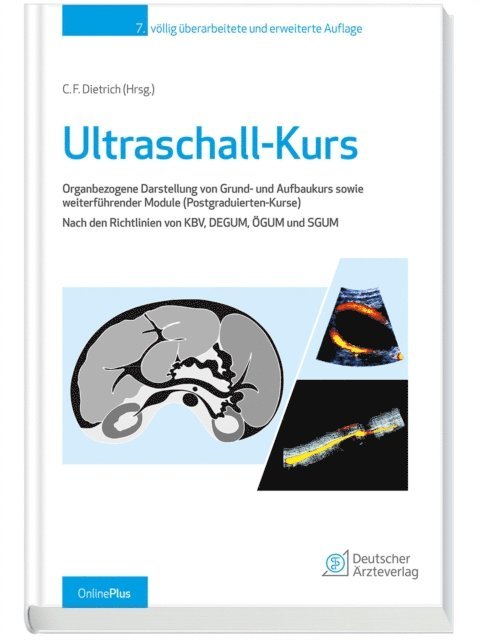
Ultraschall komplett – damit Sie sicher befundenMit der Neuauflage dieses Kursbuchs halten Sie Schritt mit den modernen Verfahren der Sonografie. Organ für Organ lernen Sie in klaren, aufschlussreichen Bildern die grundlegende sonografische Anatomie kursübergreifend kennen. Das Spektrum reicht dabei von häufigen, leicht zu erhebenden Befunden bis zu seltenen, schwerer erkennbaren Krankheitsbildern. Alle durch Richtlinien vorgegebenen Inhalte von Grund- und Aufbaukurs sowie der Module (Postgraduierten Kurse) werden berücksichtigt. Darüber hinaus eignet sich das Buch als diagnostischer Leitfaden bzw. Nachschlagewerk für den versierten Sonographeur.Neu in der 7. Auflage:- Notfallsonografie: Basisnotfallsonografie, fokussierter kardialer Ultraschall, Lungenultraschall im Notfall, fokussierter Ultraschall am Bewegungsapparat, Notfallsonografie im klinischen Kontext- Knochen- und Fraktursonografie bei Kindern- Mit OnlinePlus – die Webseite zum Buch: über 100 Videos mit Erläuterung der Untersuchungstechnik Fragen-Antwortkatalog zum Überprüfen des eigenen Wissens Tipps und Tricks alle Abbildungen des Buches Interview mit dem Herausgeber uvm.Ihre Vorteile:- Übersichtlich: organbezogene, topografische Gliederung- Komplett: Inhalt von Grund- und Aufbaukurs sowie weiterführender Spezialkurse- Strukturiert: US-Bilder mit korrespondierendem Schema- Vielfältig: B-Bild, Farb-(Power-)Doppler, Kontrastmittel, Elastografie und andere innovative Ultraschall-Methoden- Anschaulich: mit über 2.000 Abbildungen und mehr als 100 Videos- Für alle: Kursbuch und diagnostischer Leitfaden- Auf Nummer sicher: ideal für die PrüfungsvorbereitungMit dem Ultraschall-Kurs zum Sono-Profi!

Inbunden, Engelska, 2021
2 223 kr
Skickas inom 10-15 vardagar
This book aims to provide reader an overview of clinical applications of contrast-enhanced ultrasound in hepatic neoplasms diagnosis. Ultrasound images and pathological results of different hepatic neoplasms are introduced in the chapters, including benign liver tumors, malignant liver tumors, hepatic carcinoma, intrahepatic cholangiocarcinoma, rare liver benign and malignant neoplasms, regenerative nodules, inflammatory pseudotumor, parasite liver lesions, and hepatitis peliosis, etc. The combination of ultrasound findings with final histopathological results then discover the potential mechanical of contrast enhancement changes. With the development of ultrasound technology and widely application of ultrasound contrast agents (USCA) in recent decades, contrast-specific imaging modalities have been developed in combination with USCA and a low mechanical index (MI), allowing continuous real-time grey scale imaging. The updated contrast-specific software for liver diseases and hepatic tumors diagnosis has also been described described in detail. With high-resolution contrast ultrasound images during arterial phase, portal venous phase and late phase, author wants to show the whole dynamic wash-in and wash-out process of the different focal liver lesions.This book is an invaluable resource for radiologists, hepatologists and oncologists in their everyday clinical practice.
E-bok
Engelska, 20211 785 kr
Läs direkt efter köp
This book aims to provide reader an overview of clinical applications of contrast-enhanced ultrasound in hepatic neoplasms diagnosis. Ultrasound images and pathological results of different hepatic neoplasms are introduced in the chapters, including benign liver tumors, malignant liver tumors, hepatic carcinoma, intrahepatic cholangiocarcinoma, rare liver benign and malignant neoplasms, regenerative nodules, inflammatory pseudotumor, parasite liver lesions, and hepatitis peliosis, etc. The combination of ultrasound findings with final histopathological results then discover the potential mechanical of contrast enhancement changes. With the development of ultrasound technology and widely application of ultrasound contrast agents (USCA) in recent decades, contrast-specific imaging modalities have been developed in combination with USCA and a low mechanical index (MI), allowing continuous real-time grey scale imaging. The updated contrast-specific software for liver diseases and hepatic tumors diagnosis has also been described described in detail. With high-resolution contrast ultrasound images during arterial phase, portal venous phase and late phase, author wants to show the whole dynamic wash-in and wash-out process of the different focal liver lesions.This book is an invaluable resource for radiologists, hepatologists and oncologists in their everyday clinical practice.
Häftad, Engelska, 2022
1 447 kr
Skickas inom 10-15 vardagar
This book aims to provide reader an overview of clinical applications of contrast-enhanced ultrasound in hepatic neoplasms diagnosis. Ultrasound images and pathological results of different hepatic neoplasms are introduced in the chapters, including benign liver tumors, malignant liver tumors, hepatic carcinoma, intrahepatic cholangiocarcinoma, rare liver benign and malignant neoplasms, regenerative nodules, inflammatory pseudotumor, parasite liver lesions, and hepatitis peliosis, etc. The combination of ultrasound findings with final histopathological results then discover the potential mechanical of contrast enhancement changes. With the development of ultrasound technology and widely application of ultrasound contrast agents (USCA) in recent decades, contrast-specific imaging modalities have been developed in combination with USCA and a low mechanical index (MI), allowing continuous real-time grey scale imaging. The updated contrast-specific software for liver diseases and hepatic tumors diagnosis has also been described described in detail. With high-resolution contrast ultrasound images during arterial phase, portal venous phase and late phase, author wants to show the whole dynamic wash-in and wash-out process of the different focal liver lesions.This book is an invaluable resource for radiologists, hepatologists and oncologists in their everyday clinical practice.