
Farhood Saremi – författare
2 185 kr
Läs direkt efter köp

2 264 kr
Läs direkt efter köp
3 122 kr
Skickas inom 10-15 vardagar
2 860 kr
Läs direkt efter köp
2 232 kr
Skickas inom 10-15 vardagar
2 766 kr
Skickas inom 5-8 vardagar
2 766 kr
Skickas inom 5-8 vardagar
2 833 kr
Skickas inom 5-8 vardagar

3 376 kr
Läs direkt efter köp
Unique anatomic atlas provides an indispensable virtual desk dissection experience
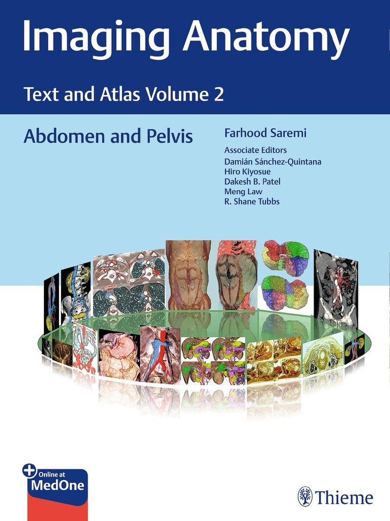
Normal imaging anatomy and variants, including diagnostic and surgical anatomy, are the cornerstones of radiologic knowledge. Imaging Anatomy: Text and Atlas Volume 2, Abdomen and Pelvis is the second in a series of four richly illustrated radiologic references edited by distinguished radiologist Farhood Saremi. The atlas is coedited by esteemed colleagues Damián Sánchez-Quintana, Hiro Kiyosue, Dakshesh B. Patel, Meng Law, and R. Shane Tubbs with contributions from an impressive cadre of international authors. Succinctly written text and superb images provide readers with a virtual, user-friendly dissection experience.
This exquisitely crafted atlas combines fundamental core anatomy principles with modern imaging and postprocessing methods to increase understanding of intricate anatomical features. Twenty-two concise chapters cover the abdominal wall, alimentary tract, liver, biliary system, pancreas, spleen, peritoneum, genitourinary system, pelvic floor, neurovasculature, and surface anatomy. Relevant anatomical components of the abdomen and pelvis are discussed, including musculature, arteries, veins, lymphatics, ducts, and innervation.
Key Highlights
High-quality cross-sectional multiplanar and volumetric color-coded CT, MRI, and angiography imaging techniques provide detailed insights on specific anatomyCross-sectional and topographic cadaveric views by internationally known anatomists coupled with more than 1,600 illustrations clearly elucidate difficult anatomical conceptsConsistently formatted chapters include an introduction, embryology, review of anatomy, discussion of anatomical variants, postsurgical anatomy, and congenital and acquired pathologiesThis unique resource provides an excellent desk reference for differentiating normal versus pathologic anatomy. It is essential reading for medical students, radiology residents and veteran radiologists, internists, and general surgeons, as well as vascular and transplant surgeons.
2 684 kr
Läs direkt efter köp
An in-depth guide to upper and lower extremity anatomy based on the latest imaging techniques
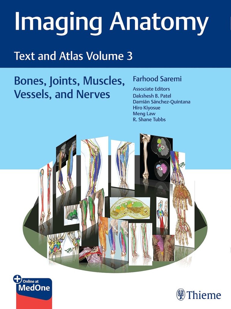
While the study of anatomy plays a fundamental role in the practice of medicine, most textbooks do not rely on modern imaging and post-processing methods to depict and increase its understanding. Imaging Anatomy: Text and Atlas Volume 3; Bones, Joints, Muscles, Vessels, and Nerves is the third in a series of four richly illustrated radiologic references edited by distinguished radiologist Farhood Saremi. The atlas is coedited by esteemed colleagues Dakshesh B. Patel, Damián Sánchez-Quintana, Hiro Kiyosue, Meng Law, and R. Shane Tubbs and features contributions from an impressive group of international experts. This book fills a gap in the literature, with descriptions of relevant anatomical components in the context of current advances in imaging technology and science.
This exquisitely crafted atlas combines fundamental core anatomy principles with modern imaging and postprocessing methods to increase understanding of intricate anatomical features. Twenty-four concise chapters cover terminology and classification of musculoskeletal structure, bones, muscles, joints, arteries, veins, nerves, and lymphatics. High-quality dissecting imaging anatomy, discussion of anatomical variants, postsurgical anatomy, and important pathology examples provide a strong foundation for differentiating normal versus pathologic anatomy.
Key Highlights
State-of-the-art CT, MR, angiography, and ultrasound techniques infused with 3D reformations, color coded volume rendering, and 3-7 Tesla MR views delineate anatomy in great detailCross-sectional and topographic cadaveric views and illustrations by world-renowned anatomists improve the ability to grasp difficult radiology conceptsConsistently formatted chapters including an introduction, embryology, review of anatomy, discussion of anatomical variants, surgical anatomy, and congenital and acquired pathologies enhance learningThis unique atlas provides a virtual, user-friendly dissection experience. It is a must-have reference for students, radiology residents, veteran radiologists, internists, general surgeons, and vascular and transplant surgeons.
3 463 kr
Läs direkt efter köp
First volume in state-of-the-art radiologic text-atlas series details anatomy of the lungs, mediastinum, and heart
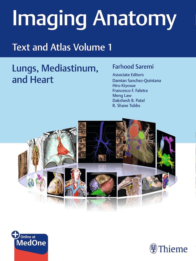
Normal imaging anatomy and variants, including both diagnostic and surgical anatomy, are the cornerstones of radiologic knowledge. Imaging Anatomy: Text and Atlas Volume 1, Lungs, Mediastinum, and Heart is the first in a series of four richly illustrated radiologic references edited by distinguished radiologist Farhood Saremi and coedited by Damian Sanchez-Quintana, Hiro Kiyosue, Francesco F. Faletra, Meng Law, Dakshesh Patel, and Shane Tubbs, with contributions from an impressive cadre of international authors.
The exquisitely crafted atlas provides high-quality multiplanar and volumetric color-coded imaging techniques utilizing CT, MRI, or angiography, supplemented by cadaveric presentations and color drawings that best elucidate each specific anatomic region. Twenty-one chapters with concise text encompass thoracic wall, mediastinum, lung, vascular, and cardiac anatomy, providing readers with a virtual dissection experience. Many anatomical variants along with pathological examples are presented.
Key Highlights
More than 600 illustrations enhance understanding of impacted regionsLung anatomy including the pleura, pulmonary arteries, pulmonary veins, and lymphaticsDiscussion of the tracheobronchial system, mediastinum and thymus, thoracic aorta and major branches, systemic veins, lymphatics and nerves of the thorax, diaphragm, and breastHeart anatomy including the atrioventricular septal region; aortic, pulmonary, mitral and tricuspid valves; coronary arteries and myocardial perfusion; coronary veins; and pericardiumThis superb resource is essential reading for medical students, radiology residents and veteran radiologists, cardiologists, as well as cardiovascular and thoracic surgeons. It provides an excellent desk reference and practical guide for differentiating normal versus pathologic anatomy.

1 495 kr
Tillfälligt slut