
Francesco S. Pavone – författare
1 021 kr
Skickas inom 10-15 vardagar
935 kr
Läs direkt efter köp
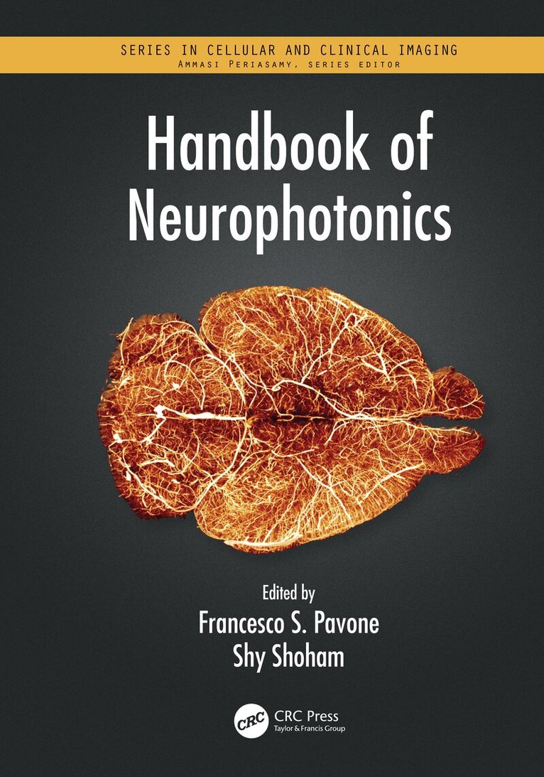
The Handbook of Neurophotonics provides a dedicated overview of neurophotonics, covering the use of advanced optical technologies to record, stimulate, and control the activity of the brain, yielding new insight and advantages over conventional tools due to the adaptability and non-invasive nature of light.
Including 32 colour figures, this book addresses functional studies of neurovascular signaling, metabolism, electrical excitation, and hemodynamics, as well as clinical applications for imaging and manipulating brain structure and function. The unifying theme throughout is not only to highlight the technology, but to show how these novel methods are becoming critical to breakthroughs that will lead to advances in our ability to manage and treat human diseases of the brain.
Key Features:
Provides the first dedicated book on state-of-the-art optical techniques for sensing and imaging across at the cellular, molecular, network, and whole brain levels. Highlights how the methods are used for measurement, control, and tracking of molecular events in live neuronal cells, both in basic research and clinical practice. Covers the entire spectrum of approaches, from optogenetics to functional methods, photostimulation, optical dissection, multiscale imaging, microscopy, and structural imaging. Includes chapters that show use of voltage-sensitive dye imaging, hemodynamic imaging, multiphoton imaging, temporal multiplexing, multiplane microscopy, optoacoustic imaging, near-infrared spectroscopy, and miniature neuroimaging devices to track cortical brain activity.
790 kr
Skickas inom 10-15 vardagar
1 247 kr
Läs direkt efter köp
2 907 kr
Skickas inom 5-8 vardagar
1 203 kr
Läs direkt efter köp
4 089 kr
Skickas inom 10-15 vardagar

935 kr
Läs direkt efter köp
The Handbook of Neurophotonics provides a dedicated overview of neurophotonics, covering the use of advanced optical technologies to record, stimulate, and control the activity of the brain, yielding new insight and advantages over conventional tools due to the adaptability and non-invasive nature of light.
Including 32 colour figures, this book addresses functional studies of neurovascular signaling, metabolism, electrical excitation, and hemodynamics, as well as clinical applications for imaging and manipulating brain structure and function. The unifying theme throughout is not only to highlight the technology, but to show how these novel methods are becoming critical to breakthroughs that will lead to advances in our ability to manage and treat human diseases of the brain.
Key Features:
Provides the first dedicated book on state-of-the-art optical techniques for sensing and imaging across at the cellular, molecular, network, and whole brain levels. Highlights how the methods are used for measurement, control, and tracking of molecular events in live neuronal cells, both in basic research and clinical practice. Covers the entire spectrum of approaches, from optogenetics to functional methods, photostimulation, optical dissection, multiscale imaging, microscopy, and structural imaging. Includes chapters that show use of voltage-sensitive dye imaging, hemodynamic imaging, multiphoton imaging, temporal multiplexing, multiplane microscopy, optoacoustic imaging, near-infrared spectroscopy, and miniature neuroimaging devices to track cortical brain activity.
1 787 kr
Läs direkt efter köp
1 725 kr
Läs direkt efter köp