G. Szikla – författare
Visar alla böcker från författaren G. Szikla. Handla med fri frakt och snabb leverans.
5 produkter
5 produkter
Häftad, Engelska, 1980
565 kr
Skickas inom 10-15 vardagar
Thus instead of being synonymous with specific procedures per- formed at the level of the basal ganglia, stereotaxis is in fact a general diagnostic and therapeutic concept, which has as its aim the precise three-dimensional representation of the patient's brain in its entirety. This three-dimensional representation is based on anatomical, neuro- radiological and other localizing information, the spatial integration Prof. Jean Talairach with friends and coworkers at the congress dinner in the Royaumont Abbey near Paris. (From left to right: Drs. ]. Bancaud, N. T. Zervas, B. Nashold, ]. Talairach, G. Szikla, F. Mundinger, P. Tournoux, ]. Peeker) of which allows a more precise "anatomical" approach to the human brain and to the surgical management of localized pathologic proces- ses. Literally, stereotaxis means "orientation in space". Taken in this general sense, all surgical procedures obviously should be stereo- tactic, at least in their principle!Two of the main themes of our meeting, namely the topic of the first day, "Surgery of Epilepsy" and that of the second "Stereotactic Cerebral Irradiation" of small brain tumours correspond to this evolution toward a global "whole brain" concept of stereotaxis. The same philosophy inspired the scientific efforts of the stereotactic group Introduction 3 of the Sainte Anne Hospital, under the leadership of its promoter, Professor Jean Talairach. This is the reason why the present Meeting is dedicated in honor of his scientific work.
Häftad, Engelska, 1984
565 kr
Skickas inom 10-15 vardagar
Local interstitial radiation therapy of intrinsic and inaccessible brain tumors is confronted with two major problems: 1. The tolerance of normal brain tissue, which is usually involved in local tumor irradiation, i.e. perifocal white matter edema and demyelinating effects, is crucial. 2. Data on radiation effects of implanted radioactive sources on neoplastic tissue, i.e. data on the radiosensitivity and on the biology of tumors, are still widely unavailable. In clinical practice the dose with which to achieve a given volume of tumor necrosis is roughly estimated. This report deals with the experimental findings of Iodine-125 and Iridium-I92 permanent implants in healthy and neoplastic brain tissue. Our own experimental findings are correlated with experimental data on Yttnul11-l)O irradiation from the literature. The study is directed to collect data on the tolerance non-tumoral brain tissue and to attempt to establish a dose-response relationship. Morphologic Changes in the Dog Brain Following Interstitial Iodine-125 Irradiation Iodine-I25 seeds (manufactured by 3 M Company, St. Paul, M. N.) with an activity of 3.55 mCi were permanently implanted under stereotactic conditions into the subcortical white matter of the gyrus coronalis of the left hemisphere in 6 beagle dogs. One animal with a non-radioactive seed implanted under similar operative conditions served as a control. The animals were allowed to survive 25, 46, 71, 94, 248, 368 days, respectively, after the implantation. The control animal survived 46 days. A detailed description of the experimental procedure is given elsewhere 16. 17.
E-bok
PDF, Engelska, 20121 174 kr
Läs direkt efter köp
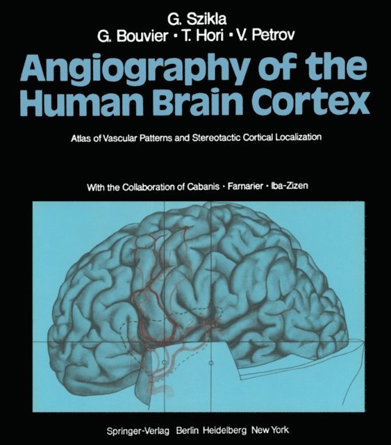
Considering the numerous works dealing with the angiography of the human brain, the book presented by SZIKLA et al. might seem to some to be devoted to superfluous precision, especially as it is inspired by "stereotactic" thinking. The large arterial trunks and their branches were described by anatomists for a long time, then were restudied by neuroradiologists for recognition in a more and more detailed manner on arteriograms. However, until now no encompassing work has been done to specify precisely the relationship of the blood vessels to that large and important organ, the human brain cortex, thereby permitting the recognition of the sulci and gyri as a function of the successive curves imposed on the various vessels by the deep infoldings of the cortex. Insofar as the radiologic evaluation of the cerebral cortex is concerned, fractional pneumoencephalography allows the injection of a number of sulci and fissures via the subarachnoid spaces. It should be pointed out, however, that sufficiently complete and interpretable images are obtained only under favorable circumstances (successful technique, cerebral atrophy, absence of cerebral edema, absence of arachnoid symphysis, etc. ). In addition a large number of sulci cannot be made visible by pneumography for strictly anatomic reasons such as the level of their opening into cisternal spaces.
E-bok
PDF, Engelska, 2012734 kr
Läs direkt efter köp
E-bok
PDF, Engelska, 2012708 kr
Läs direkt efter köp
Local interstitial radiation therapy of intrinsic and inaccessible brain tumors is confronted with two major problems: 1. The tolerance of normal brain tissue, which is usually involved in local tumor irradiation, i.e. perifocal white matter edema and demyelinating effects, is crucial. 2. Data on radiation effects of implanted radioactive sources on neoplastic tissue, i.e. data on the radiosensitivity and on the biology of tumors, are still widely unavailable. In clinical practice the dose with which to achieve a given volume of tumor necrosis is roughly estimated. This report deals with the experimental findings of Iodine-125 and Iridium-I92 permanent implants in healthy and neoplastic brain tissue. Our own experimental findings are correlated with experimental data on Yttnul11-l)O irradiation from the literature. The study is directed to collect data on the tolerance non-tumoral brain tissue and to attempt to establish a dose-response relationship. Morphologic Changes in the Dog Brain Following Interstitial Iodine-125 Irradiation Iodine-I25 seeds (manufactured by 3 M Company, St. Paul, M. N.) with an activity of 3.55 mCi were permanently implanted under stereotactic conditions into the subcortical white matter of the gyrus coronalis of the left hemisphere in 6 beagle dogs. One animal with a non-radioactive seed implanted under similar operative conditions served as a control. The animals were allowed to survive 25, 46, 71, 94, 248, 368 days, respectively, after the implantation. The control animal survived 46 days. A detailed description of the experimental procedure is given elsewhere 16. 17.