
L. Jeanmart – författare
Visar alla böcker från författaren . Handla med fri frakt och snabb leverans.
6 produkter
6 produkter

Häftad, Engelska, 1977
1 115 kr
Skickas inom 10-15 vardagar
In this book are published papers presented at the first meeting about tomodensito- metry (Computer Tomography) which the CEPUR organized in Luxembourg in March 1977. CEPUR (College d'enseignement post-universitaire de Radiologie) is an international medical association having as its main aim the promotion of courses in advanced radiology. Several sections deal with the subspecializations, one of which is Com- puter Tomography. Thanks to the fruitful cooperation of several University Hospitals (Ancona, Leuven, Montpellier, Bruxelles, Strasbourg), the two-day meeting organized by Dr. Capesius in Luxembourg covered a certain number of aspects of clinical tomodensitometry in the brain as well as in the trunk. We hope that this volume will be the first of a series dealing with the actual problems in clinical radiology. Leuven/Bruxelles/Strasbourg A. BAERT L. JEANMART A. WACKENHEIM Contents I. Introduction to the Technology of Computer Tomography K. Ungerer. With 18 Figures ...2 II. Head Sellar Region: Normal and Pathologic Conditions. U. Salvolini, F. Menichelli, and U. Pasquini. With 40 Figures ...14 Empty Sella and Pituitary Gland. J.L. Dietemann and A. Wackenheim. Wi th 3 Figures .* . . * . *. *...* . ., 38 Midline Lesions. D. Baleriaux-Waha, L.L. Mortelmans, M. Dupont, and L. Jeanmart. With 17 Figures ...*.. 39 Ventriculocisternal Pathology in Children. D. Touitou. With 9 Figures ...* . . * ...47 Endocranial Calcifications. J.H. Vandresse, G. Cornelis, and A.
E-bok
PDF, Engelska, 20121 132 kr
Läs direkt efter köp
E-bok
PDF, Engelska, 20121 132 kr
Läs direkt efter köp
It was our aim to place at the disposal of radiologists within a short time an atlas of high-quality, valuable pictures of abdominal CT without We felt that, the image degradation inherent to slower scanning apparatus. notwithstanding rapid evolution in CT scanning apparatus and the resulting rapid advances in our clinical knowledge about the value, limitations and applications of this new diagnostic imaging modality, an effort should be made to realize an atlas of reference. From more than 7,000 patients studied with abdominal CT, we tried to assemble images with maximal anatomical detail, which implies the use of large window settings, being well aware that in daily routine practice basic CT methodology includes the use of different window settings for optimal information. It was, however, a conscious decision not to use comparative images with large and small window settings in order to stay within a reasonable total number of figures. Much emphasis has been placed upon the use of contrast enhancement by intravenous contrast media. This is based on the conviction that essen tially new and better morphological information about normal and pathological processes within the abdomen can be obtained because short exposure times now allow one to capture the rapidly changing aspect and degree of contrast enhancement of the lesions as a function of their vascu larity.
E-bok
PDF, Engelska, 20121 174 kr
Läs direkt efter köp
The purpose of this book is to provide the radiologist with information which is "as practical as possible" for the everyday use of computerized tomography (CT) in the field of cervical, thoracic, and musculoskeletal pathology. The approach is simple. For each region the following information is pre sented: (1) a general schematic introduction, summarizing the main indications for CT and its specific usefulness; (2) a series of pictures of normal structures with a precise and practical identification; and (3) a selection of pictures of pathological structures, with a description and a short comment, aimed at covering the largest possible field of CT indications and interests. This approach has been applied to the following areas: cervical pathology, with one section dealing with the larynx and hypopharynx; the thorax, specifi cally to pulmonary diseases, pleural and parietal pathology, and the mediasti num, with special sections dealing with tumours, the heart, and large vessels; the spine, which is of growing importance in clinical CT; and finally the pathol ogy of the musculoskeletal system in general, with special attention being paid to the developing field of orthopaedic CT measuring methods.
E-bok
PDF, Engelska, 20121 408 kr
Läs direkt efter köp
E-bok
PDF, Engelska, 20121 174 kr
Läs direkt efter köp
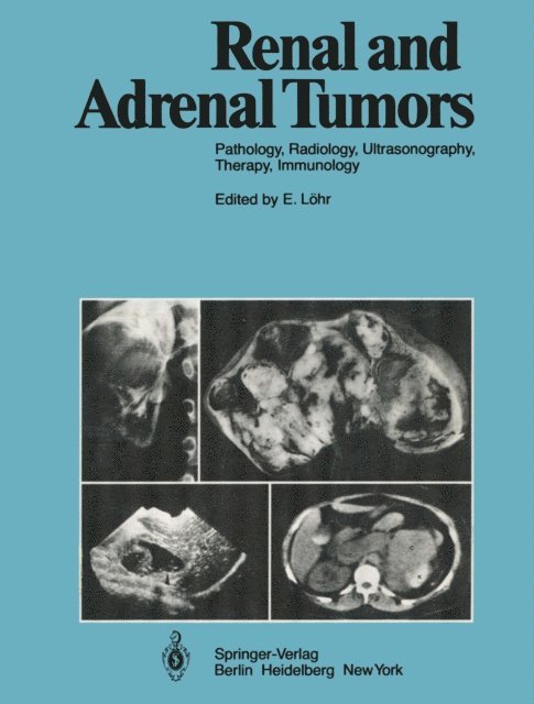
The present volume constitutes an attempt to compile contem porary features of diagnosis and treatment of renal and adrenal tumors. A thorough survey of the field is ensured by the authors'' considerable scientific experience. Tumors of the kidneys and the adrenal glands are being diagnosed and treated by physicians of different medical disciplines. For both types of tumor, the pathologic cellular substrate is of crucial importance in diagnosis and therapy. In recent years significant diagnostic advances have been made, ranging from angiography through ultrasonography and computer tomography to immunology. New impulses in oncologic therapy have occurred in surgery, radiation therapy, and tumor emboliza tion. A further important topic is renal tumors in infants. Such tumors involve special aspects of both diagnosis and therapy and also have a distinctive prognosis. We are indebted both to Springer-Verlag, who supported us in our intention to write this book, and to our colleagues, whose help is greatly appreciated. For the authors: E. LOHR Essen/Heidelberg, September 1979 Contents (Chapters marked with an asterisk have been translated by H.-U. Eickenberg) Pathology of Renal and Adrenal Neoplasms L.-D. Leder, H.J. Richter, and Chr. Stambolis ...... . 1. Tumors and Tumor-Like Lesions of the Kidney in the Adult 1.1. General Remarks . 1.2. Heterotopic Tissue . . . . l.2.l. Adrenal Tissue . . . . . .