
Lorenzo Cerroni – författare

4 986 kr
Skickas inom 5-8 vardagar
2 111 kr
Läs direkt efter köp
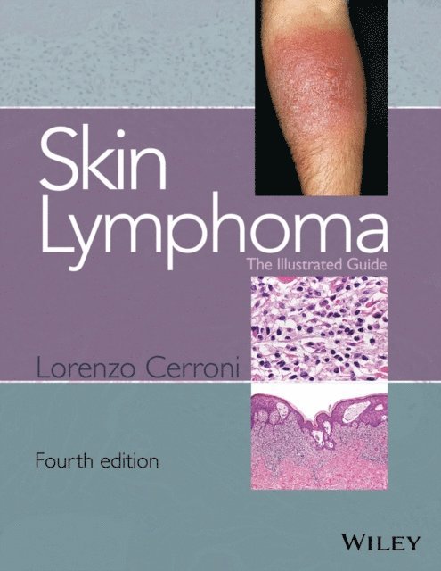
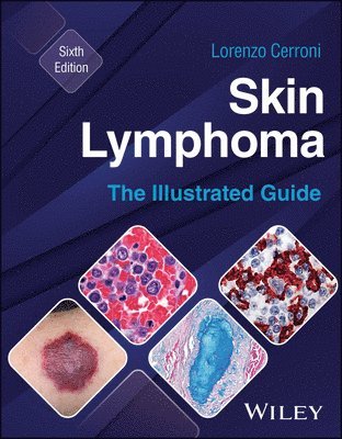
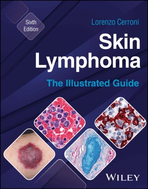
Skin lymphomas are relatively rare but potentially fatal
They can easily be misdiagnosed as benign skin diseases. Dermatologists and pathologists need to have a good understanding of the clinical presentations and the pathological correlates of this challenging disease to ensure the correct diagnosis and most appropriate treatment is provided.
Skin Lymphoma: The Illustrated Guide is a full-color illustrated text and atlas that combines pathology with clinical features and treatment. Jam-packed with pictures that illustrate key diagnostic points and supplemented by teaching cases to highlight effective decision making. It includes all newly identified entities and provides pointers on how to differentiate these. In addition, it also covers all inflammatory conditions that may simulate skin lymphomas (cutaneous pseudolymphomas).
Skin Lymphoma: The Illustrated Guide provides
Clinical and pathological guidance on diagnosis of skin lymphomas Clear illustrations to aid visual recognition Nomenclature according to WHO/EORTC classifications Cases to enhance the scope for teaching and learning
This fully refreshed fourth edition continues to provide the clinical excellence that has helped a generation of dermatologists and dermatopathologists to effectively diagnose skin lymphoma.

2 337 kr
Skickas inom 5-8 vardagar
2 688 kr
Läs direkt efter köp
2 594 kr
Läs direkt efter köp
3 671 kr
Skickas inom 5-8 vardagar

4 066 kr
Läs direkt efter köp
554 kr
Läs direkt efter köp
5 709 kr
Skickas inom 10-15 vardagar

7 067 kr
Läs direkt efter köp
Die 2. Auflage dieses umfassenden Lehr- und Nachschlagewerks enthält alles, was man über die moderne Dermatohistopathologie wissen muss. Das gesamte Buch wurde von einem erfahrenen Autorenteam komplett überarbeitet und um neue Diagnosen, Klassifizierungen und die ICD-O Codierung ergänzt. Die Abbildungen wurden weitestgehend erneuert und ihre Zahl erhöht.
Alle Hauterkrankungen und deren wichtigsten Varianten werden in Definition, Klinik, Histologie und Differenzialdiagnose vorgestellt. Besonders die zahlreichen hervorragenden histologischen Farbabbildungen zeigen, worauf es bei der histopathologischen Diagnose ankommt.
Die Techniken der Dermatohistopathologie wie Immunhistochemie, Antigen-Mapping oder Molekularpathologie werden praxisnah dargestellt. Es findet sich nun auch ein Kapitel über konfokale Laser-Scanning-Mikroskopie.
Für alle Dermatologen und Pathologen, die bereits dermatopathologisch arbeiten oder noch lernen wollen, wie man histologische Befunde der Haut interpretiert.
Die Herausgeber
Univ.-Prof. Dr.med. Lorenzo Cerroni, Universitätsklinik für Dermatologie und Venerologie, LKH-Universitätsklinikum Graz, Leiter der Forschungseinheit für Dermatopathologie
Prof. Dr. med. Claus Garbe, Hautklinik, Universitätsklinikum Tübingen, Sektionsleiter Dermatologische Onkologie, und Sprecher des Hauttumorzentrums
Prof. Dr. med. Dieter Metze, Klinik für Hautkrankheiten, Universitätsklinikum Münster, Oberarzt und Leiter der Dermatohist
ologiePD Dr. med. Heinz Kutzner, Dermatopathologie Friedrichshafen
Prof. em. Dr. med. Helmut Kerl, Universitätsklinik für Dermatologie und Venerologie, LKH-Universitätsklinikum Graz
3 541 kr
Läs direkt efter köp