Orazio Schillaci – författare
Visar alla böcker från författaren Orazio Schillaci. Handla med fri frakt och snabb leverans.
13 produkter
Atlas of Hybrid Imaging Sectional Anatomy for PET/CT, PET/MRI and SPECT/CT Vol. 1: Brain and Neck
Sectional Anatomy for PET/CT, PET/MRI and SPECT/CT
Av Mario Leporace, Ferdinando Calabria m. fl.
Häftad, 2023
1818 kr
Lägg i varukorg
For the first time, in this atlas nuclear physicians and radiologists cover the entire hybrid nuclear medicine (PET/CT, SPECT/CT and PET/MRI), based on their own case studies. The structure in three volumes represents an user friendly guide for interpreting PET and SPECT in relation to …
Atlas of Hybrid Imaging Sectional Anatomy for PET/CT, PET/MRI and SPECT/CT Vol. 1: Brain and Neck
Sectional Anatomy for PET/CT, PET/MRI and SPECT/CT
Av Antonio Bagnato, Alfonso Ciaccio m. fl.
E-bok, 2023
2329 kr
Lägg i varukorg
For the first time, in this atlas nuclear physicians and radiologists cover the entire hybrid nuclear medicine (PET/CT, SPECT/CT and PET/MRI), based on their own case studies.The structure in three volumes represents an user friendly guide for interpreting PET and SPECT in relation to co-registered …
Atlas of Hybrid Imaging Sectional Anatomy for PET/CT, PET/MRI and SPECT/CT Vol. 2: Thorax Abdomen and Pelvis
Sectional Anatomy for PET/CT, PET/MRI and SPECT/CT
Av Mario Leporace, Ferdinando Calabria m. fl.
Häftad, 2023
1672 kr
Lägg i varukorg
Atlas of Hybrid Imaging of the Thorax, Abdomen and Pelvis, Volume Two: Sectional Anatomy for PET/CT, PET/MRI and SPECT/CT provides a guide for interpreting PET and SPECT in relation to co-registered CT and/or MRI. In this atlas, exclusively dedicated to thorax, abdomen and pelvis, nuclear …
Atlas of Hybrid Imaging Sectional Anatomy for PET/CT, PET/MRI and SPECT/CT Vol. 2: Thorax Abdomen and Pelvis
Sectional Anatomy for PET/CT, PET/MRI and SPECT/CT
Av Antonio Bagnato, Alfonso Ciaccio m. fl.
E-bok, 2023
2310 kr
Lägg i varukorg
Atlas of Hybrid Imaging of the Thorax, Abdomen and Pelvis, Volume Two: Sectional Anatomy for PET/CT, PET/MRI and SPECT/CT provides a guide for interpreting PET and SPECT in relation to co-registered CT and/or MRI. In this atlas, exclusively dedicated to thorax, abdomen and pelvis, nuclear …
Atlas of Hybrid Imaging Sectional Anatomy for PET/CT, PET/MRI and SPECT/CT Vol. 3: Heart, Lymph Node and Musculoskeletal System
Sectional Anatomy for PET/CT, PET/MRI and SPECT/CT
Av Mario Leporace, Ferdinando Calabria m. fl.
Häftad, 2023
1672 kr
Lägg i varukorg
Atlas of Hybrid Imaging of the Heart, Lymph Nodes and Musculoskeletal System, Volume Three: Sectional Anatomy for PET/CT, PET/MRI and SPECT/CT provides a guide for interpreting PET and SPECT in relation to co-registered CT and/or MRI. In this atlas, exclusively dedicated to heart, lymph nodes and …
Atlas of Hybrid Imaging Sectional Anatomy for PET/CT, PET/MRI and SPECT/CT Vol. 3: Heart, Lymph Node and Musculoskeletal System
Sectional Anatomy for PET/CT, PET/MRI and SPECT/CT
Av Antonio Bagnato, Alfonso Ciaccio m. fl.
E-bok, 2023
2310 kr
Lägg i varukorg
Atlas of Hybrid Imaging of the Heart, Lymph Nodes and Musculoskeletal System, Volume Three: Sectional Anatomy for PET/CT, PET/MRI and SPECT/CT provides a guide for interpreting PET and SPECT in relation to co-registered CT and/or MRI. In this atlas, exclusively dedicated to heart, lymph nodes and …
Radiopharmaceuticals
A Guide to PET/CT and PET/MRI
Av Orazio Schillaci, Ferdinando Calabria
E-bok, 2019
1232 kr
Lägg i varukorg
This book provides a rapid and concise guide to PET (PET/CT and PET/MRI) molecular imaging, concentrating extensive information on PET radiopharmaceuticals in a single volume. The book reflects the rapid development of several PET tracers over the last decade, as a result of which the “traditional” …
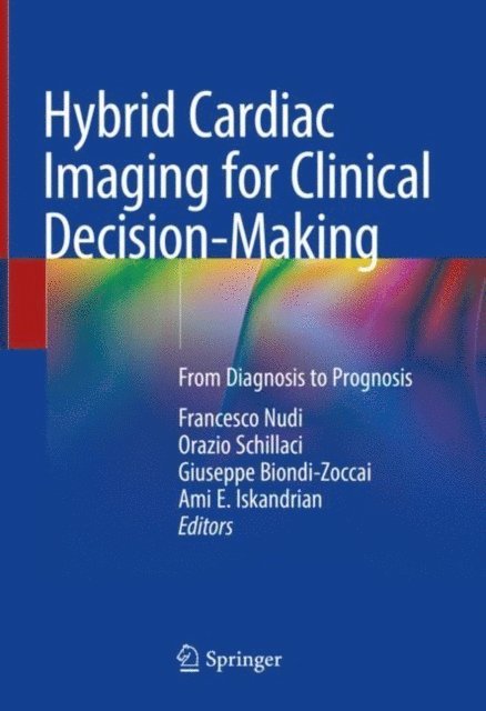
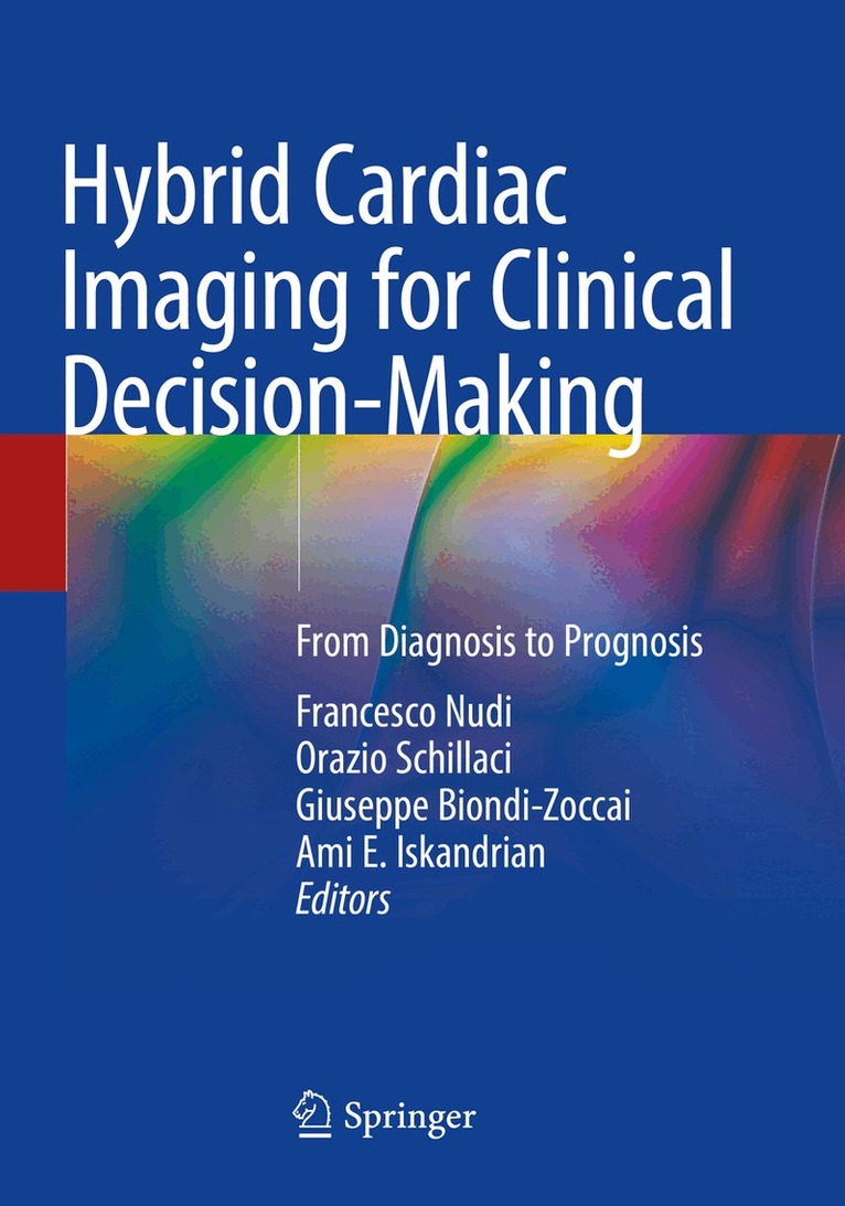
Hybrid Cardiac Imaging for Clinical Decision-Making
From Diagnosis to Prognosis
Av Francesco Nudi, Orazio Schillaci m. fl.
Inbunden, 2022
1690 kr
Lägg i varukorg
Performing any diagnostic test in medicine is always a matter of trying to get the condition of the patient diagnosed properly with the least effort, exposure, discomfort and at the same time with the lowest possible error probability.Pre-test probability is helpful but often imprecise, effectively …
Hybrid Cardiac Imaging for Clinical Decision-Making
From Diagnosis to Prognosis
Av Ami E. Iskandrian, Giuseppe Biondi-Zoccai m. fl.
E-bok, 2022
1634 kr
Lägg i varukorg
Performing any diagnostic test in medicine is always a matter of trying to get the condition of the patient diagnosed properly with the least effort, exposure, discomfort and at the same time with the lowest possible error probability. Pre-test probability is helpful but often imprecise, …
Hybrid Cardiac Imaging for Clinical Decision-Making
From Diagnosis to Prognosis
Av Francesco Nudi, Orazio Schillaci m. fl.
Häftad, 2023
1241 kr
Lägg i varukorg
Performing any diagnostic test in medicine is always a matter of trying to get the condition of the patient diagnosed properly with the least effort, exposure, discomfort and at the same time with the lowest possible error probability.Pre-test probability is helpful but often imprecise, effectively …
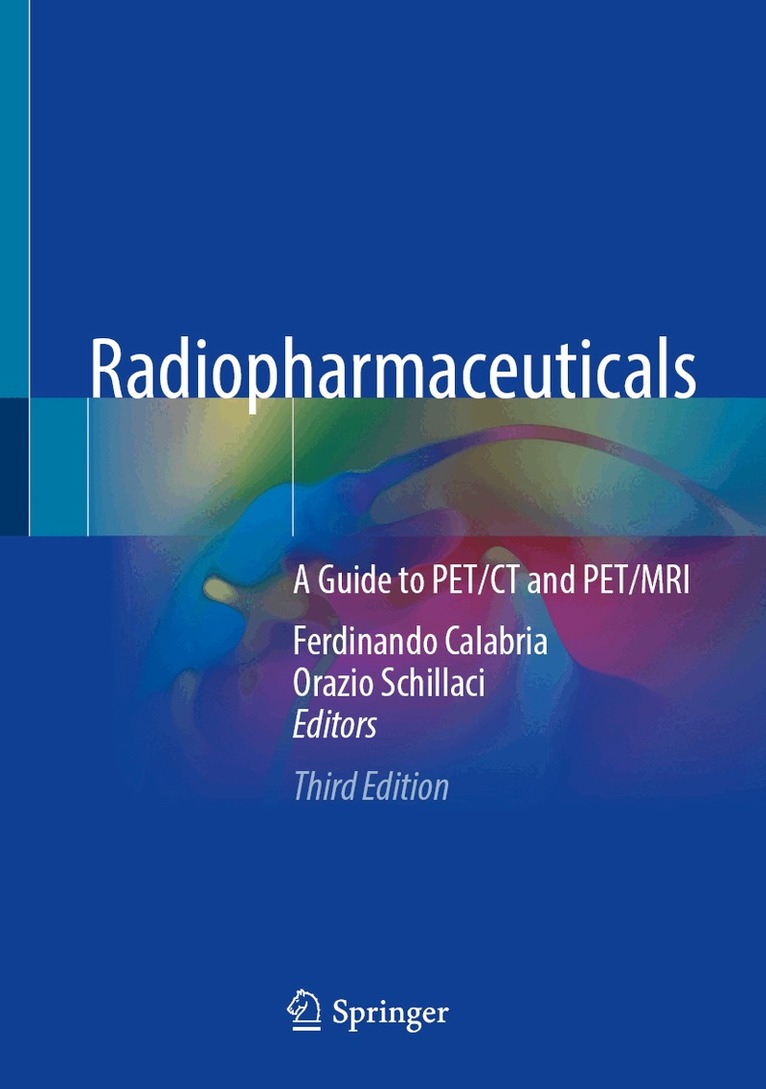
Radiopharmaceuticals
A Guide to PET/CT and PET/MRI
Av Ferdinando Calabria, Orazio Schillaci
Inbunden, 2024
1577 kr
Lägg i varukorg
The book is a practical and concise guide to PET tracers actually useful for PET/CT and PET/MR imaging.Gaining knowledge on radiopharmaceuticals is often difficult, since their features can be only found in single original journal articles.This book aims to present to the reader, in one single …
Radiopharmaceuticals
A Guide to PET/CT and PET/MRI
Av Orazio Schillaci, Ferdinando Calabria
E-bok, 2024
2014 kr
Lägg i varukorg
The book is a practical and concise guide to PET tracers actually useful for PET/CT and PET/MR imaging. Gaining knowledge on radiopharmaceuticals is often difficult, since their features can be only found in single original journal articles.This book aims to present to the reader, in one single …
Radiopharmaceuticals
A Guide to PET/CT and PET/MRI
Av Ferdinando Calabria, Orazio Schillaci
Häftad, 2025
1129 kr
Lägg i varukorg
The book is a practical and concise guide to PET tracers actually useful for PET/CT and PET/MR imaging.Gaining knowledge on radiopharmaceuticals is often difficult, since their features can be only found in single original journal articles.This book aims to present to the reader, in one single …