
Paul Babyn – författare
982 kr
Skickas inom 10-15 vardagar
1 134 kr
Skickas inom 5-8 vardagar
1 612 kr
Skickas inom 11-20 vardagar
1 039 kr
Läs direkt efter köp
125 cases addressing "real-life" clinical problems
Complete with the insights of leading pediatric radiologists, Teaching Atlas of Pediatric Imaging provides 125 cases that address the challenging "real-life" clinical problems that you are likely to encounter. Each chapter presents a different case with a complete patient work-up that includes clinical presentation, diagnosis, differential diagnoses, radiological and clinical findings, treatment summary and suggested readings. With a view to providing the opportunity for self-assessment, the authors omit the diagnosis from the first pages of each case to enable self-testing and review.
Highlights:
Easy-to-access arrangement of cases based on anatomy: head and neck, chest, heart, abdomen, pelvis, and the musculoskeletal system Coverage of a wide spectrum of diseases, from the very common to more important uncommon entities, including congenital heart disease, bone dysplasias and more Differential diagnoses for each case, as well as information on etiology, pathology, treatment, and complications "Pearls" and "Pitfalls" that help you identify important points and avoid errors in image interpretationHere is a valuable resource for the clinician at every level, from the resident preparing for the radiology board examinations, to the practitioner seeking the Certificate of Added Qualification in Pediatric Radiology, to the general radiologist or pediatrician seeking a practical reference text.
1 576 kr
Läs direkt efter köp
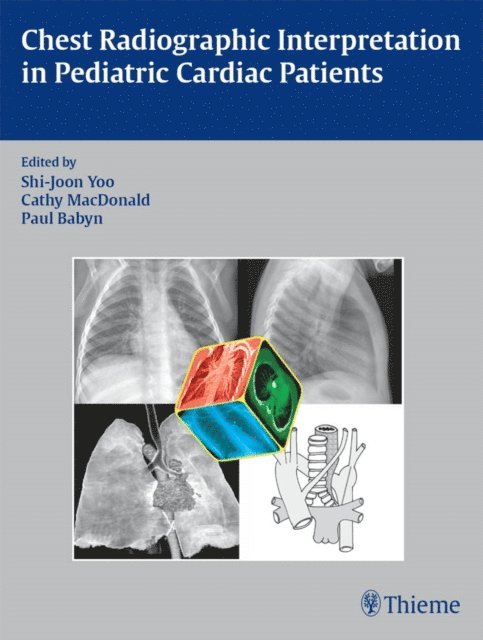
Beautifully illustrated case-based guide to reading chest radiographs in pediatric cardiac patients
From congenital or acquired problems in newborns to cardiovascular abnormalities in older children, this comprehensive text explains how to interpret chest radiographs and how to report that information in day-to-day practice. It steers readers through this often challenging area using numerous practical case examples, more than 400 high-quality radiographs, drawings and specimen photographs, straightforward explanations of findings, and the collective experience of several of the world''s foremost experts on cardiac imaging and pediatric cardiology. After an introductory discussion of normal cardiac anatomy and imaging, the authors provide readers with a systematic approach to understanding chest images in children with congenital or acquired heart disease followed by useful bulleted synopses of basic pathologic features, clinical manifestations and radiographic findings.
While the trend in recent decades has been toward increasingly sophisticated imaging modalities, this book successfully illustrates that there is still an extraordinary amount of diagnostic and therapeutic information to be found in chest x-rays.
Features
Multiple perspectives from imagers, cardiologists, and cardiac surgeons on the pathology of congenital heart disease High-resolution radiographs, detailed drawings, and specimen photographs vividly elucidate interpretative principles Summary of pediatric cardiovascular surgical procedures provides context and practical examples of what to expect when viewing post-operative chest radiographsThis book will improve the accuracy and confidence of any radiologist, cardiologist, or clinician involved in the interpretation of pediatric chest images and is ideal for residents and fellows in radiology and pediatric cardiology.