Rudolf Nieuwenhuys - Böcker
Visar alla böcker från författaren Rudolf Nieuwenhuys. Handla med fri frakt och snabb leverans.
6 produkter
6 produkter
2 499 kr
Skickas inom 10-15 vardagar
This book demonstrates that the systematic study of gene expression patterns in embryonic and adult brains, in combination with selected data from earlier studies, can pave the way for a new neuromorphology, the most salient features of which may be summarized as follows: (1) Causal analysis of molecular patterning at neural plate and early neural tube stages has shown that the CNS is essentially organized into transverse neural segments or neuromeres and longitudinal zones which follow the curved axis of the brain. (2) The FMUs initially represent thin neuroepithelial fields; in the course of further development they are transformed into three-dimensional radial units, extending from the ventricular surface to the meningeal surface of the brain. (3) The principal histogenetic processes, including cellular proliferation, cell migration and differentiation, essentially take place within the confines of these radial units, controlled by characteristic sets of developmental regulatory genes. (4) Although most developing neurons migrate radially and settle within their own FMU, at many locations neuroblasts leave the FMU where they were produced and migrate tangentially to other nearby or remote territories, colonizing parts of foreign FMUs. (5) Many structural complexes in the adult brain, including the cerebral and cerebellar cortices, are the products of radial and tangential intermingling of migrated cell contingents.(6) By using appropriate molecular markers, all neuron types in the adult CNS can be traced back to a specific progenitor zone within a specific FMU, and the progeny of any FMU can be traced to their final positions with the help of selective labeling approaches. (7) Early outgrowing axons form bundles, which tend to pass close to the border zones of the radial units. By means of their molecularly diversely tuned growth cones, these extending axons decide how to behave at each boundary they encounter, sometimes even reorienting at right angles. Collectively these early axonal bundles form a checkerboard-like scaffold, which accentuates the molecular regionalization of the CNS and leads to the formation of topographically ordered synaptic fields. The book covers all of these aspects in detail, providing a morphologic model (blueprint) that highlights the natural coordinates of CNS structure resulting from the conserved molecularly controlled shaping phenomena within morphogenetic fields.
1 524 kr
Skickas inom 7-10 vardagar
Includes chapters that introduce illustrations of the gross anatomy, the blood supply and the microstructure of the central nervous system and deal with the development, topography and functional anatomy of the spinal cord, brain stem and cerebellum, diencephalons and telencephalon.
1 414 kr
Skickas inom 10-15 vardagar
This comprehensive reference is clearly destined to become the definitive anatomical basis for all neuroscience research. The book provides a complete overview and comparison of the structural organization of all vertebrate groups, ranging from amphioxus and lamprey through fishes, amphibians and birds to mammals. The large specialised section of the work, devoted to the CNS of the various vertebrate groups, is preceded by introductory chapters on neurons, cell masses, fibre tracts, morphogenesis, methodology, and techniques. Although focusing on structure, the authors provide functional correlations throughout. This monumental work is, and will remain, unique; the only source of such brilliant illustrations at both the macroscopic and microscopic levels.
1 527 kr
Skickas inom 10-15 vardagar
The present edition of The Human Central Nervous System differs considerably from its predecessors. In previous editions, the text was essentially confined to a section dealing with the various functional systems of the brain. This section, which has been rewritten and updated, is now preceded by 15 newly written chapters, which introduce the pictorial material of the gross anatomy, the blood vessels and meninges and the microstructure of its various parts and deal with the development, topography and functional anatomy of the spinal cord, the brain stem and the cerebellum, the diencephalon and the telencephalon. Great pains have been taken to cover the most recent concepts and data. As suggested by the front cover, there is a focus on the evolutionary development of the human brain. Throughout the text numerous correlations with neuropathology and clinical n- rology have been made. After much thought, we decided to replace the full Latin terminology, cherished in all previous editions, with English and Anglicized Latin terms. It has been an emotional farewell from beautiful terms such as decussatio hipposideriformis W- nekinkii and pontes grisei caudatolenticulares. Not only the text, but also the p- torial material has been extended and brought into harmony with the present state of knowledge. More than 230 new illustrations have been added and many others have been revised. The number of macroscopical sections through the brain has been extended considerably. Together, these illustrations now comprise a complete and convenient atlas for interpreting neuroimaging studies.
982 kr
Skickas inom 10-15 vardagar
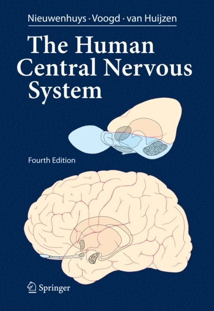
The Human Central Nervous System di Nieuwenhuys è ormai un "classico" nel campo della neurologia. Le eccezionali illustrazioni che arricchiscono il testo rappresentano ancora oggi la sua principale caratteristica: 390 tra figure e disegni costituiscono un atlante completo e pratico per l’interpretazione degli studi di neuroimaging. La seconda edizione italiana è basata sulla quarta edizione in lingua inglese, che è stata estensivamente aggiornata e implementata: quindici nuovi capitoli illustrano l’anatomia macroscopica, l’apporto ematico e la microstruttura del sistema nervoso centrale, e descrivono lo sviluppo, la topografia e l’anatomia funzionale del midollo spinale, del tronco dell’encefalo, del cervelletto, del diencefalo e del telencefalo.