
Tim Leiner – författare
Visar alla böcker från författaren . Handla med fri frakt och snabb leverans.
5 produkter
5 produkter
Inbunden, Engelska, 2022
1 890 kr
Skickas inom 10-15 vardagar
This book provides an overview of current and potential applications of artificial intelligence (AI) for cardiothoracic imaging. Most AI systems used in medical imaging are data-driven and based on supervised machine learning. Clinicians and AI specialists can contribute to the development of an AI system in different ways, focusing on their respective strengths. Unfortunately, communication between these two sides is far from fluent and, from time to time, they speak completely different languages. Mutual understanding and collaboration are imperative because the medical system is based on physicians’ ability to take well-informed decisions and convey their reasoning to colleagues and patients.This book offers unique insights and informative chapters on the use of AI for cardiothoracic imaging from both the technical and clinical perspective. It is also a single comprehensive source that provides a complete overview of the entire process of the development and use of AI in clinical practice for cardiothoracic imaging. The book contains chapters focused on cardiac and thoracic applications as well more general topics on the potentials and pitfalls of AI in medical imaging. Separate chapters will discuss the valorization, regulations surrounding AI, cost-effectiveness, and future perspective for different countries and continents. This book is an ideal guide for clinicians (radiologists, cardiologists etc.) interested in working with AI, whether in a research setting developing new AI applications or in a clinical setting using AI algorithms in clinical practice. The book also provides clinical insights and overviews for AI specialists who want to develop clinically relevant AI applications.
E-bok
Engelska, 20221 785 kr
Läs direkt efter köp
This book provides an overview of current and potential applications of artificial intelligence (AI) for cardiothoracic imaging. Most AI systems used in medical imaging are data-driven and based on supervised machine learning. Clinicians and AI specialists can contribute to the development of an AI system in different ways, focusing on their respective strengths. Unfortunately, communication between these two sides is far from fluent and, from time to time, they speak completely different languages. Mutual understanding and collaboration are imperative because the medical system is based on physicians’ ability to take well-informed decisions and convey their reasoning to colleagues and patients.This book offers unique insights and informative chapters on the use of AI for cardiothoracic imaging from both the technical and clinical perspective. It is also a single comprehensive source that provides a complete overview of the entire process of the development and use of AI in clinical practice for cardiothoracic imaging. The book contains chapters focused on cardiac and thoracic applications as well more general topics on the potentials and pitfalls of AI in medical imaging. Separate chapters will discuss the valorization, regulations surrounding AI, cost-effectiveness, and future perspective for different countries and continents. This book is an ideal guide for clinicians (radiologists, cardiologists etc.) interested in working with AI, whether in a research setting developing new AI applications or in a clinical setting using AI algorithms in clinical practice. The book also provides clinical insights and overviews for AI specialists who want to develop clinically relevant AI applications.
Häftad, Engelska, 2022
1 336 kr
Skickas inom 10-15 vardagar
This book provides an overview of current and potential applications of artificial intelligence (AI) for cardiothoracic imaging. Most AI systems used in medical imaging are data-driven and based on supervised machine learning. Clinicians and AI specialists can contribute to the development of an AI system in different ways, focusing on their respective strengths. Unfortunately, communication between these two sides is far from fluent and, from time to time, they speak completely different languages. Mutual understanding and collaboration are imperative because the medical system is based on physicians’ ability to take well-informed decisions and convey their reasoning to colleagues and patients.This book offers unique insights and informative chapters on the use of AI for cardiothoracic imaging from both the technical and clinical perspective. It is also a single comprehensive source that provides a complete overview of the entire process of the development and use of AI in clinical practice for cardiothoracic imaging. The book contains chapters focused on cardiac and thoracic applications as well more general topics on the potentials and pitfalls of AI in medical imaging. Separate chapters will discuss the valorization, regulations surrounding AI, cost-effectiveness, and future perspective for different countries and continents. This book is an ideal guide for clinicians (radiologists, cardiologists etc.) interested in working with AI, whether in a research setting developing new AI applications or in a clinical setting using AI algorithms in clinical practice. The book also provides clinical insights and overviews for AI specialists who want to develop clinically relevant AI applications.
Inbunden, Engelska, 2008
262 kr
Skickas inom 10-15 vardagar
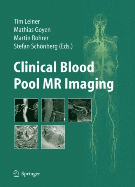
Magnetic resonance angiography has made great strides, with continuing improvements in hardware, pulse sequencing, and know-how allowing ever-increasing speed, resolution, and suppression of artifacts. However, an inherent physical barrier has always been limited SNR. Gadolinium contrast agents help to increase SNR by facilitating T1 relaxation, but they can be injected only at a finite rate and at a limited molar dose, and there is a rapid drop in c- centration following the brief arterial phase due to redistribution into the extracellular fluid compartment. With its sixfold increase in T1 relaxivity, blood pool distribution, and longer serum half-life, Vasovist® represents a new breakthrough which promises to revolutionize MRA image quality once again. This excellent treatise on Vasovist®, created by a team of exceptional faculty who are pioneers in MR angiography, covers the basic techniques, safety, efficacy, image processing, and pharmacoeconomic details, to successfully implement a new level of MRA image quality with this new contrast agent. In addition to improving all the usual arterial phase MRA - plications, the blood pool distribution opens up new possibilities, including detecting internal bleeding and imaging stent graft endoleaks, which are reviewed in detail. In the complex, competitive field of cardiovascular imaging, this book articulates the cutting edge in imaging vascular disease.
E-bok
PDF, Engelska, 2009326 kr
Läs direkt efter köp
Magnetic resonance angiography has made great strides, with continuing improvements in hardware, pulse sequencing, and know-how allowing ever-increasing speed, resolution, and suppression of artifacts. However, an inherent physical barrier has always been limited SNR. Gadolinium contrast agents help to increase SNR by facilitating T1 relaxation, but they can be injected only at a finite rate and at a limited molar dose, and there is a rapid drop in c- centration following the brief arterial phase due to redistribution into the extracellular fluid compartment. With its sixfold increase in T1 relaxivity, blood pool distribution, and longer serum half-life, Vasovist® represents a new breakthrough which promises to revolutionize MRA image quality once again. This excellent treatise on Vasovist®, created by a team of exceptional faculty who are pioneers in MR angiography, covers the basic techniques, safety, efficacy, image processing, and pharmacoeconomic details, to successfully implement a new level of MRA image quality with this new contrast agent. In addition to improving all the usual arterial phase MRA - plications, the blood pool distribution opens up new possibilities, including detecting internal bleeding and imaging stent graft endoleaks, which are reviewed in detail. In the complex, competitive field of cardiovascular imaging, this book articulates the cutting edge in imaging vascular disease.