
Tommaso Scarabino – författare
1 669 kr
Skickas inom 10-15 vardagar
1 622 kr
Läs direkt efter köp
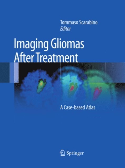
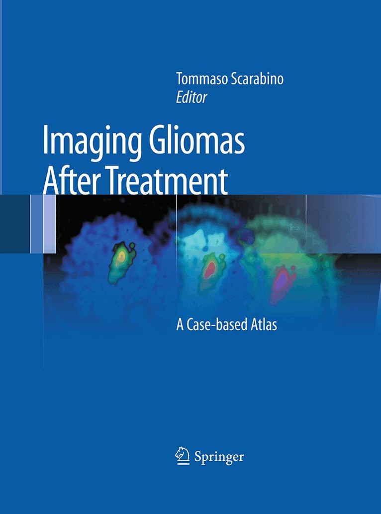
This book illustrates the characteristics of imaging after treatment in brain gliomas. It describes in detail the modifications to brain tissue, both healthy and pathological, that can manifest after surgery, radiotherapy or chemotherapy treatment. These modifications are discussed in terms of both how they occur in the immediate post-treatment period, and in the long term. The imaging methods used include CT with and without the addition of contrast medium, but above all MRI, which involves the use of routine basic sequences and mainly advanced study techniques such as diffusion, perfusion, spectroscopy and cortical activation. The aim of the text is to equip neuroradiologists with adequate expertise in post-treatment examinations reporting, allowing them to perform an effective differential diagnosis between the persistency or recurrence of illness and the effects of short or long-term treatment.
The book is divided into a general section, which addresses the classification of cerebral tumors, the surgical treatment options, radiotherapy and chemotherapy protocols; and a section on clinical cases that employs rich iconography, making it quick and easy to consult.
This second edition has been updated to reflect the new WHO classification system from 2016; new surgical, radiotherapy and chemotherapeutic treatment options; and (in the iconography section) the new sequences available from the manufacturers of RM scanners.
1 226 kr
Skickas inom 10-15 vardagar
1 558 kr
Skickas inom 10-15 vardagar
1 459 kr
Läs direkt efter köp
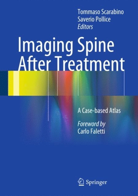
This atlas illustrates the characteristics of imaging after surgical spine treatment. The previous edition has been thoroughly updated and new surgical treatment options are presented. Furthermore, all clinical cases feature new images with the new sequences available from the manufacturers of Magnetic Resonance scanners.
The imaging methods presented in the book are MRI, involving the use of routine basic sequences and advanced study techniques, and CT with and without administration of contrast medium.
The modifications of the spine, both healthy and pathological, that can occur immediately after surgical treatment and in the long term, are described in detail.Atlas contents are organized in a general part, with the classification of spine pathology and the surgical treatment options, and a part with clinical cases enriched by a wealth of images.
This easy-to-consult publication addresses neuroradiologists who wish to gain an adequate expertise in post-treatment examinations reporting in order to be able to perform an effective differential diagnosis.
1 115 kr
Skickas inom 10-15 vardagar
1 336 kr
Skickas inom 10-15 vardagar
1 785 kr
Läs direkt efter köp
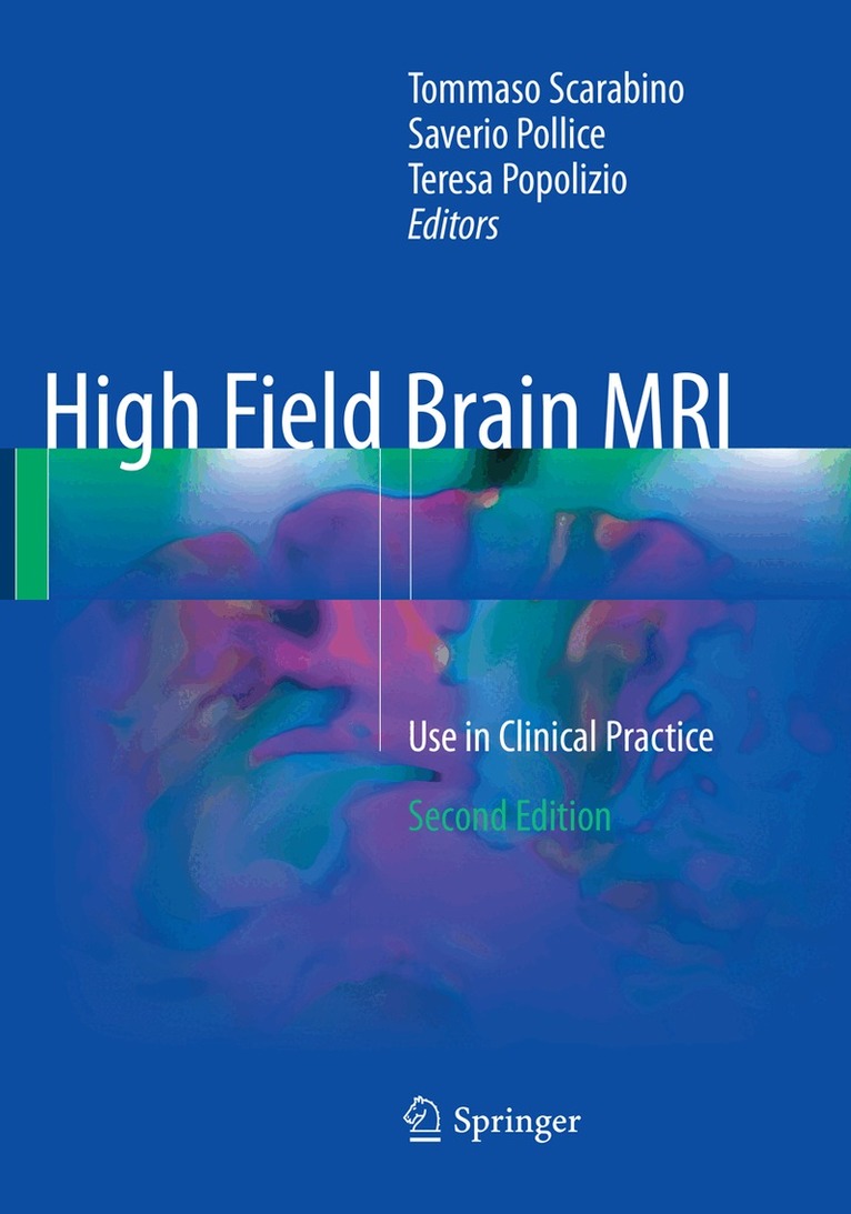
This richly illustrated book summarizes the state of the art in brain MRI with the 3-Tesla high-field scanner. The aim is to clarify the numerous advantages of using a 3-T high magnetic field MR scanner, especially in terms of sensitivity and specificity. The first section describes techniques for standard MR examination of the brain. Full descriptions are then provided of advanced protocols such as MR angiography, diffusion-weighted imaging (DWI), perfusion-weighted imaging (PWI), MR spectroscopy, MR tractography, and functional imaging. Differences in diagnostic features when performing these examinations at 3 T and at 1.5 T are highlighted. In addition, safety issues relating to the installation and use of a high-field scanner are discussed. The second section then describes and illustrates in detail each of the main clinical applications of 3-T MRI in the human brain: trauma, stroke, white matter disease, Parkinson’s disease, Alzheimer’s disease, brain tumors, inflammatory disease, psychiatric disorders, etc. A final chapter is dedicated to the evaluation of 7-T MRI within both research and potential clinical settings. The book will be a valuable tool for general radiologists, neuroradiologists, trainees, and technicians.
1 336 kr
Skickas inom 10-15 vardagar

1 890 kr
Skickas inom 10-15 vardagar
2 436 kr
Läs direkt efter köp
1 890 kr
Skickas inom 10-15 vardagar
539 kr
Skickas inom 10-15 vardagar
571 kr
Läs direkt efter köp
1 467 kr
Läs direkt efter köp
1 622 kr
Läs direkt efter köp
This book reviews in detail the role of neuroradiological imaging in the evaluation of patients who have undergone surgery or interventional radiology procedures, and particularly its value in the documentation of normal and pathological post-treatment changes, detection of complications, and follow-up.
The opening sections describe pretreatment images in various conditions, including trauma, degenerative disc disease, and osteoporosis, and the different types of neurosurgical and interventional treatment that may be used. The post-treatment appearances of normal sequelae and complications on conventional radiography, CT, and MRI are then documented in detail on the basis of a large series of clinical cases, with a wealth of images. Guidance is provided on selection of one or a combination of imaging modalities. This book will be an invaluable clinical and research tool not only for neuroradiologists but also for neurosurgeons, and interventional radiologists.
1 157 kr
Skickas inom 11-20 vardagar