
Torsten Bert Möller – författare
638 kr
Skickas inom 3-6 vardagar

638 kr
Skickas inom 3-6 vardagar
686 kr
Skickas inom 3-6 vardagar

658 kr
Skickas inom 5-8 vardagar
926 kr
Skickas inom 3-6 vardagar
318 kr
Skickas inom 5-8 vardagar
303 kr
Skickas inom 5-8 vardagar

953 kr
Skickas inom 3-6 vardagar
2 269 kr
Skickas inom 5-8 vardagar
866 kr
Läs direkt efter köp

2 533 kr
Skickas inom 3-6 vardagar

2 533 kr
Skickas inom 3-6 vardagar

2 533 kr
Skickas inom 3-6 vardagar
953 kr
Skickas inom 3-6 vardagar
822 kr
Skickas inom 3-6 vardagar
752 kr
Skickas inom 3-6 vardagar
809 kr
Skickas inom 3-6 vardagar

686 kr
Skickas inom 3-6 vardagar

982 kr
Skickas inom 3-6 vardagar
554 kr
Läs direkt efter köp
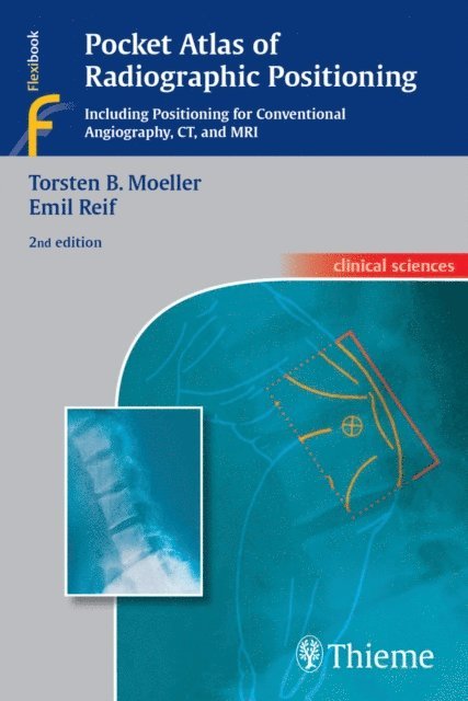
Radiographic positioning - comprehensive and concise
Now in its second edition, Pocket Atlas of Radiographic Positioning is a practical how-to guide that provides the detailed information you need to reproducibly obtain high-quality radiographic images for optimal evaluation and interpretation of normal, abnormal, and pathological anatomic findings. It shows positioning techniques for all standard examinations in conventional radiology, with and without contrast, as well as basic positioning for CT and MRI. For each type of study a double-page spread features an exemplary radiograph, positioning sketches, and helpful information on imaging technique and parameters, criteria for the best radiographic view, and patient preparation. Clearly organized to be used in day-to-day practice, the atlas serves as an ideal companion to Moeller and Reif''s Pocket Atlas of Radiographic Anatomy and their three-volume Pocket Atlas of Cross-Sectional Anatomy.
Highlights of the second edition:
New chapters on positioning in MRI and CT, including multislice CT A greatly expanded section on mammography Special features, including information on the advantages of a specific view, variations of positions, and practical tips and tricks Nearly 500 excellent radiographs and drawings demonstrating the relationship between correct patient positioning and effective diagnostic imagesPocket Atlas of Radiographic Positioning, Second Edition is an excellent desk or pocket reference for radiologists, radiology residents, and for radiologic technologists.
554 kr
Läs direkt efter köp
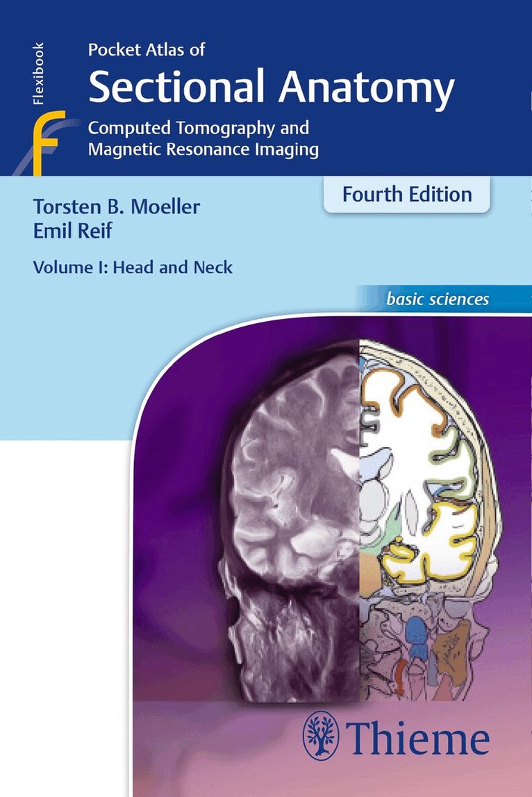
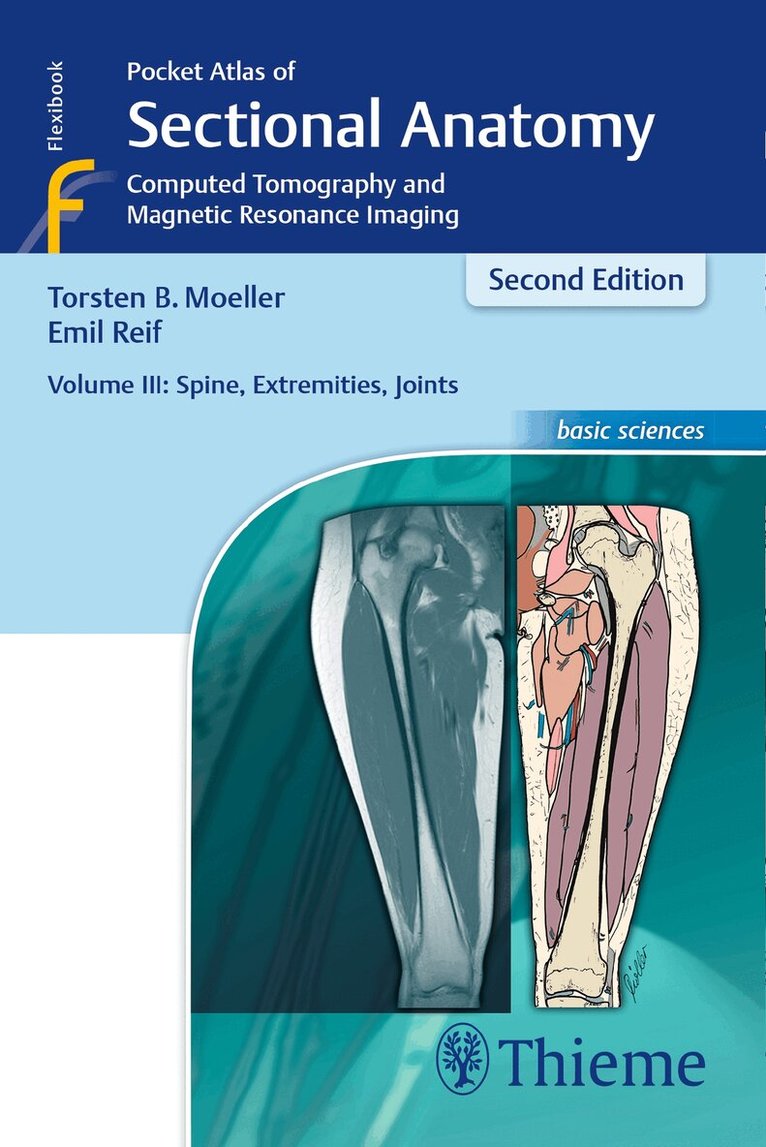
This comprehensive, easy-to-consult pocket atlas is renowned for its superb illustrations and ability to depict sectional anatomy in every plane. Together with its two companion volumes, it provides a highly specialized navigational tool for all clinicians who need to master radiologic anatomy and accurately interpret CT and MR images.
Special features of Pocket Atlas of Sectional Anatomy:
Didactic organization in two-page units, with high-quality radiographs on one side and brilliant, full-color diagrams on the other Hundreds of high-resolution CT and MR images made with the latest generation of scanners (e.g., 3T MRI, 64-slice CT) Color-coded schematic drawings that indicate the level of each section Consistent color coding, making it easy to identify similar structures across several slicesUpdates for the 4th edition of Volume II:
CT imaging of the chest and abdomen in all 3 planes: axial, sagittal, and coronal New back-cover foldout featuring pulmonary and hepatic segments and lymph node stations Follows standard international classifications of the American Heart Association for cardiac vessels and the AJCC/UICC for mediastinal lymph nodesCompact, easy-to-use, highly visual, and designed for quick recall, this book is ideal for use in both the clinical and study settings.
554 kr
Läs direkt efter köp
The method of making findings and the systematic method of interpreting images are still relevant today.
This book deals with a subject that is seemingly trivial: normal findings in radiology. However, the normal is also the frequent, but not always the simple. Every radiologist has experienced difficulties in the systematics of imaging and with phrasing the diagnosis. This book is intended to answer three questions:
How should the diagnosis be phrased? Which general scheme can I use to evaluate an image; how can I check for the degree of normality? Which data allow me to evidence normality, and what measurements should be taken?In spite of the introduction of digital X-ray imaging, the interpretation of conventional X-rays has not basically changed during the past ten years, which is evidenced by the development of this book. Since its first publication in 1987, it has gone through several unchanged reprints. In the 2nd German edition the text has hardly been changed; however, several new images have been added.
The method of making findings and the systematic method of interpreting images are still relevant today. Consequently, the strict and systematic organization of the book was left unchanged. The new layout leads to a clearer accessibility of the material for the reader.
554 kr
Läs direkt efter köp
The key for any beginning radiologist who wishes to recognize pathological findings is to first acquire an ability to distinguish them from normal ones. This outstanding guide gives beginning radiologists the tools they need to systematically approach and recognize normal MR and CT images.
Highlights include:
Reference-quality images from the author''s own teaching files show all standard normal findings as seen in CT and MRIChecklists in each section offer the reader a systematic way to approach the imagesThorough guidelines to help beginning radiologists dictate their reportsLists of standard measurements and tips for ruling out pathology