
Val M. Runge – författare
1 222 kr
Läs direkt efter köp
1 076 kr
Skickas inom 5-8 vardagar
1 558 kr
Läs direkt efter köp
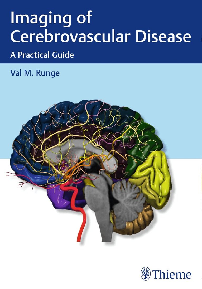
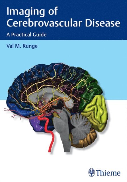
Imaging plays an integral role in the diagnosis and intervention of debilitating, often fatal vascular diseases of the brain, such as ischemic and hemorrhagic stroke, aneurysms, and arteriovenous malformations (AVMs). Written by a world renowned neuro-radiologist and pioneer in the early adoption of magnetic resonance (MR) technology, Imaging of Cerebrovascular Disease is a concise yet remarkably thorough textbook that advances the reader''s expertise on this subject.
The text draws on the author''s vast personal experience, case studies, and traditional educational sources, offering didactic dialogue with accompanying images. A practical clinical resource organized into six chapters, this book offers unparalleled breadth in delineating the diagnostic and treatment usages of modern imaging techniques. Chapter one sets a foundation with extensive coverage of modalities and their applications, including MR, computed tomography (CT) and digital subtraction angiography (DSA). Subsequent chapters cover utilization of imaging techniques specific to underlying pathologies.
Key Features:
In-depth discussion of medical and neuroradiologic/neurosurgical interventions, focusing on the use of imaging prior to, during, and following treatmentComprehensive text enhanced with more than 700 high-quality imagesDetailed evaluation of normal brain anatomy, as well as gyral anatomy in brain ischemia, an important subtopicAdvantages, disadvantages, mortality, and morbidity of surgery (clipping) versus endovascular techniques (coiling and flow diversion) for aneurysmsPresented in a style that facilitates cover-to-cover reading, this is an essential tool for residents and fellows, and provides a robust study guide prior to sitting for relevant certification exams. It is also a quick, invaluable reference for radiologists, neurosurgeons, and neurologists in the midst of a busy clinical day.
641 kr
Läs direkt efter köp
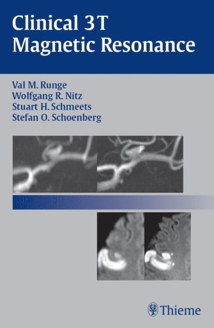
An introduction to the premier clinical imaging field strength for MR
Here is the first textbook to present a practical overview of the basic principles and clinical applications for 3 tesla (3 T) MR imaging. Organized into sections according to anatomical location, each case study is presented in a concise, two-page unit that enables the reader to digest and review the material in small sections. The author describes the situations that dictate the use of 3 T and explains the numerous clinical advantages of this field strength by drawing comparisons to corresponding studies at 1.5 T.
Highlights:
Case studies from leading international experts covering the breadth of clinical MR Recommendations for how to optimize image quality and how to interpret the clinical findings Easy-to-follow descriptions of the strengths and limitations of 3 T 400 high-quality clinical images and illustrations depicting key concepts Discussion of the various pulse sequence approachesClinical 3T Magnetic Resonance is essential reading for all radiologists, radiology residents, MR physicists, and MR technologists seeking to master this emerging diagnostic tool.
797 kr
Läs direkt efter köp
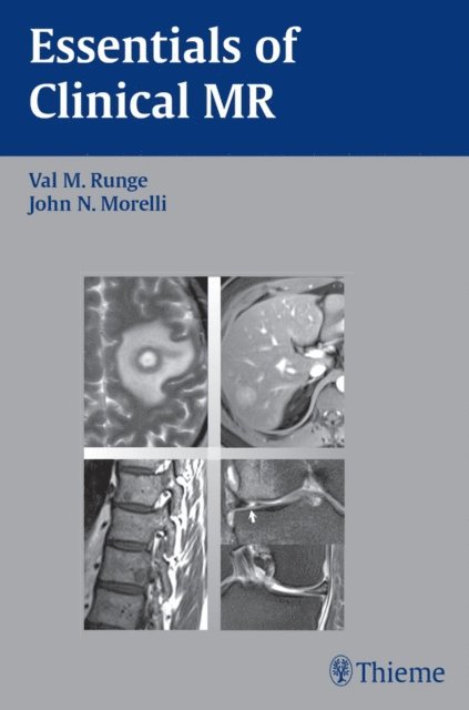
Gain crucial diagnostic skills with this image-rich guide to clinical MR interpretation
Essentials of Clinical MR provides readers with the must-have background they need to read magnetic resonance images and make successful clinical diagnoses.
Intuitively arranged by body region, this user-friendly manual explores the most commonly encountered diseases through concise case examples supplemented by clearly labeled MRI images. Using the case descriptions as starting points, each section thoroughly surveys a different anatomic area and provides tips on imaging techniques followed by an in-depth discussion of the image interpretation.
Features
Complete coverage of the diseases most frequently seen in the clinical practice of MRI Over 650 images clearly illustrate the MR appearance of each disease Highly relevant information on contrast media and contrast enhanced MRAThis easily accessible guide is both the ideal introductory text for radiology residents, MR technologists, and medical students, as well as a practical daily reference for anyone involved in the interpretation of clinical MRIs. Residents will find it an invaluable companion when studying for exams.
1 212 kr
Läs direkt efter köp
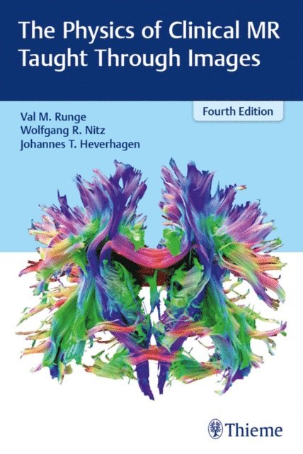
The fourth edition of The Physics of Clinical MR Taught Through Images
The Physics of Clinical MR Taught Through Images Fourth Edition by Val Runge, Wolfgang Nitz, and Johannes Heverhagen presents a unique and highly practical approach to understanding the physics of magnetic resonance imaging. Each physics topic is described in user-friendly language and accompanied by high-quality graphics and/or images. The visually rich format provides a readily accessible tool for learning, leveraging, and mastering the powerful diagnostic capabilities of MRI.
Key Features
More than 700 images, anatomical drawings, clinical tables, charts, and diagrams, including magnetization curves and pulse sequencing, facilitate acquisition of highly technical content.Eight systematically organized sections cover core topics: hardware and radiologic safety; basic image physics; basic and advanced image acquisition; flow effects; techniques specific to the brain, heart, liver, breast, and cartilage; management and reduction of artifacts; and improvements in MRI diagnostics and technologies.Cutting-edge topics including contrast-enhanced MR angiography, spectroscopy, perfusion, and advanced parallel imaging/data sparsity techniques.Discussion of groundbreaking hardware and software innovations, such as MR-PET, 7 T, interventional MR, 4D flow, CAIPIRINHA, radial acquisition, simultaneous multislice, and compressed sensing.A handy appendix provides a quick reference of acronyms, which often differ from company to company.The breadth of coverage, rich visuals, and succinct text make this manual the perfect reference for radiology residents, practicing radiologists, researchers in MR, and technologists.
1 991 kr
Läs direkt efter köp
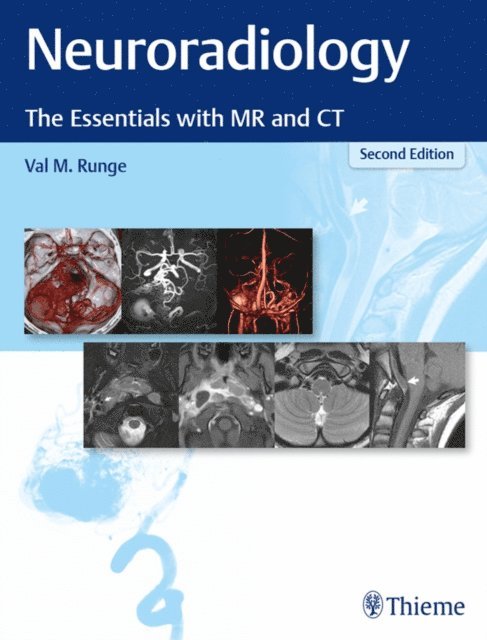
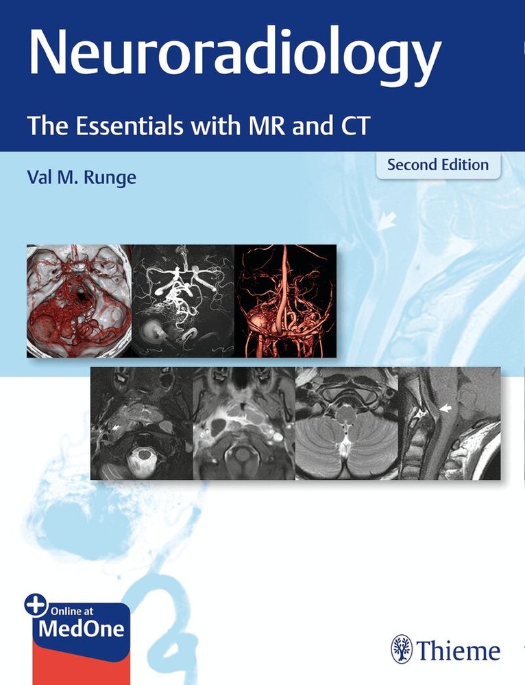
An image-rich neuroradiology reference and board prep from renowned experts
Neuroradiology: The Essentials with MR and CT, Second Edition, written by world-renowned neuroradiologist and MRI pioneer Val Runge, builds on the acclaimed prior edition. The splendidly illustrated compendium features in-depth discussion of important imaging findings, focused primarily on common disease processes. An impressive cadre of international experts contribute to the text, which is written from a clinical radiology perspective and draws from firsthand experiences. MRI physics pearls and tips throughout the book will help radiologists avoid common pitfalls.
Designed as a practical educational resource for clinical neuroradiology, the text is divided into three sections: the brain, head and neck, and spine. The brain and spine chapters are divided into subsections covering normal anatomy and major disease categories such as congenital, traumatic, degenerative, vascular, infectious, and neoplastic. Head and neck chapters are organized by major anatomic region. Clinical cases encompass the use of advanced imaging techniques such as perfusion, high-resolution imaging, and spectroscopy.
Key Features
About 1,300 high-quality MR and CT images illustrate relevant findings and cases, including those often not well-described in more traditional academic textbooksNew figures, updates on ultra-high-field 7T MRI, and additional in-depth text on cerebrovascular disease – especially brain aneurysms and AVMsCovers a wide array of diseases – from stroke and multiple sclerosis to cases one might see once a year, such as glutaric acidemia type 1 and CADASILThis excellent clinical resource provides a robust study prep for the boards and is a must-read for radiology residents prior to neuroradiology rotation. A quick reference for diagnosing challenging cases encountered in daily practice, it will also benefit neuroradiology fellows and general radiologists.
1 680 kr
Skickas inom 11-20 vardagar
1 337 kr
Skickas inom 10-15 vardagar
1 214 kr
Läs direkt efter köp
The text is organized into concise chapters, each discussing an important point relevant to clinical MR and illustrated largely with images from routine patient exams. The topics covered encompass the breadth of thefield, from imaging basics and pulse sequences to advanced topics including contrast-enhanced MR angiography, spectroscopy, perfusion and advanced parallel imaging/data sparsity techniques. Discussion of the latest hardware and software innovations, for example next generation low field MR, deep learning, MR-PET, 7 T, interventional MR, 4D flow, CAIPIRINHA, spiral techniques, radial acquisition, simultaneous multislice, compressed sensing and MR fingerprinting, is included because these topics are critical to current clinical practice as well as to future advances. Included in the fifth edition are a large number of new topics, keeping the text up to date in this increasingly complex field. The text has also been thoroughly revised to include additional relevant clinical images, to improve the clarity of descriptions, and to increase the depth of content.
The book is highly recommended for radiologists, physicists, and technologists interested in the background of image acquisition used in standard as well as specialized clinical settings.
949 kr
Skickas inom 10-15 vardagar