
Vishali Gupta – författare
Visar alla böcker från författaren . Handla med fri frakt och snabb leverans.
4 produkter
4 produkter
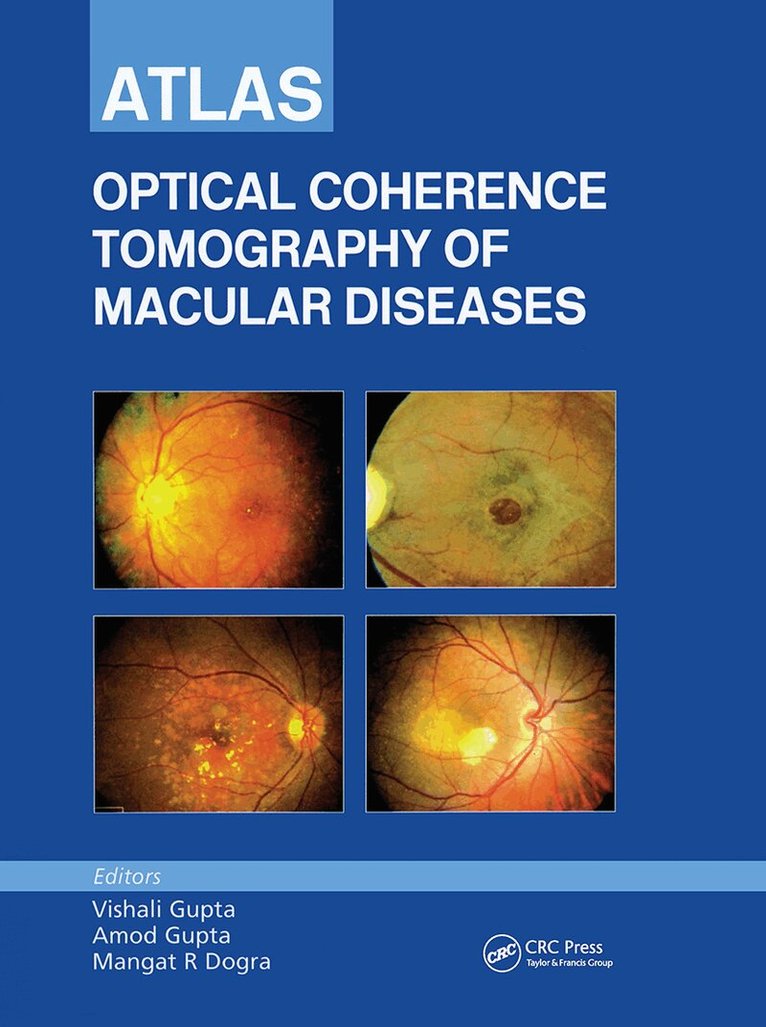
Inbunden, Engelska, 2004
2 734 kr
Skickas inom 10-15 vardagar
The emergence of Optical Coherence Tomography (OCT) in recent years has revolutionized the way we see the retina. Providing, in real time, high-resolution cross-sectional images of the macula that are very similar to obtaining in vivo histopathological specimens, OCT represents a major advance in the diagnostics of retinal disease. The excitement of working with this new tool has been dampened by the non-availability of any standard textbook on the subject and meant that every new finding on the OCT saw us rushing to the library almost on a daily basis to locate any published reports on the subject. Until now.Containing nearly 900 scans of both normal and diseased appearances, most in full color, Atlas of Optical Coherence Tomography of Macular Diseases covers how to use Stratus OCT for diagnosing various macular disorders, identifying correct therapeutic approaches and monitoring the responses to therapies and interventions. The authors provide brief case summaries, fundus photographs, fluorescein angiography, and the OCT images and the follow up images. They discuss OCT applications for diagnosis, management, and follow-up in diabetic macular edema, macular hole, taut posterior hyaloid membrane, vitreofoveal traction, idiopathic central serous chorioretinoplasty, submacular pathology, and more.
Inbunden, Engelska, 2019
8 310 kr
Skickas inom 10-15 vardagar
Uveitis comprises a group of intraocular diseases that poses a diagnostic challenge to the ophthalmologists of all subspecialties, including uveitis specialists. The morphological expression of the diseases may be characteristic at times, but it is challenging to detect and recognize subtle clinical clues that might indicate towards a particular etiology: infectious, non-infectious or masquerade. Ancillary investigations including fluorescein angiography, indocyanine green angiography, auto fluorescence imaging, ultrasound, ultrasound biomicroscopy, laser flaremeter and optical coherence tomography play a crucial role in reaching a specific diagnosis and require proper interpretation in context of each disease variety. In addition, the radiological imaging, systemic work-up and laboratory investigations aid in establishing the diagnosis. This is a first comprehensive atlas that compiles these varied findings in each type of uveitis. It describes all uveitic entities employing the aid of slit lamp and anterior segment photographs, fundus photographs, ancillary investigations where applicable and relevant laboratory and radiologic investigations. The case-based format sets out the context clearly to match the diagnostic findings with the treatment plan. Covered in 136 chapters and more than 1000 images, this atlas is an essential reference guide for all uveitis specialists and general ophthalmologists. Regular updates on new insights, techniques and treatments meet the point-of-need requirements of the busy readers looking for most current information.
E-bok
Engelska, 201910 591 kr
Läs direkt efter köp
Uveitis comprises a group of intraocular diseases that poses a diagnostic challenge to the ophthalmologists of all subspecialties, including uveitis specialists. The morphological expression of the diseases may be characteristic at times, but it is challenging to detect and recognize subtle clinical clues that might indicate towards a particular etiology: infectious, non-infectious or masquerade. Ancillary investigations including fluorescein angiography, indocyanine green angiography, auto fluorescence imaging, ultrasound, ultrasound biomicroscopy, laser flaremeter and optical coherence tomography play a crucial role in reaching a specific diagnosis and require proper interpretation in context of each disease variety. In addition, the radiological imaging, systemic work-up and laboratory investigations aid in establishing the diagnosis. This is a first comprehensive atlas that compiles these varied findings in each type of uveitis. It describes all uveitic entities employing the aid of slit lamp and anterior segment photographs, fundus photographs, ancillary investigations where applicable and relevant laboratory and radiologic investigations. The case-based format sets out the context clearly to match the diagnostic findings with the treatment plan. Covered in 136 chapters and more than 1000 images, this atlas is an essential reference guide for all uveitis specialists and general ophthalmologists. Regular updates on new insights, techniques and treatments meet the point-of-need requirements of the busy readers looking for most current information.
Inbunden, Engelska, 2009
4 751 kr
Tillfälligt slut