
Electron Microscopy in Biology and Medicine – serie
Visar alla böcker i serien Electron Microscopy in Biology and Medicine. Handla med fri frakt och snabb leverans.
15 produkter
15 produkter
Inbunden, Engelska, 1990
3 331 kr
Skickas inom 10-15 vardagar
The calcified tissues have fundamental functions in the biology of organisms, not only because their strength, solidity, and elasticity permit movement and mechanical activities, and protect soft tissues against traumatic forces, but also on account of their role in mineral homeostasis. For this reason, extensive investigation in the last 30 years has provided much to explain the complex chemical and physical processes occurring in cells and matrices composing the skeleton, and their alterations in pathological conditions. The use of ultrastructural methods such as immunocytochemistry, scanning and transmission electron microscopy, cytoautoradiography, freeze/fracture etching, high voltage, etc. has proven to be of great value when applied to cells and matrix components of bone and cartilage, in spite of the technical difficulties due to the hardness of these tissues. However, available information on this subject is disseminated in a variety of scientific and medical articles. This volume is an attempt to collect together the most significant data on the ultrastructure of cartilage and bone in normalcy and pathology. Obviously, it cannot be a complete report of all these data, its principal aim being that of: a) giving a comprehensive statement of the results concerning the basic structures common to these tissues, especially collagen fibrils, noncollagenous proteins, and proteoglycans, and their relationships with the mineral substance (for which another volume of this series can also be consulted; see Ruggeri A. , Motta P. M. (eds.
Inbunden, Engelska, 1991
3 331 kr
Skickas inom 10-15 vardagar
Inbunden, Engelska, 1992
893 kr
Skickas inom 10-15 vardagar
Recently, attention has been called to the role that microvascular organization plays in the functional morphology of all organs and tissues, both in normal and pathological conditions. Since its development by Murakami, the corrosion cast method for scanning electron microscopy has come to be considered one of the most efficient means in clarifying the three-dimensional features of the microcirculation of organs and tissues. "Scanning Electron Microscopy of Vascular Casts: Methods and Applications" was planned to supply fundamental and new information regarding microcirculation studies to general biologists, anatomists, pathologists and clinicians. The contributions to this volume contain original findings and electron micrographs obtained by using recently improved corrosion methods. The variety of papers in this book should be useful to many, and aim to provide both the basic and clinically oriented readers with ideas, suggestions, and original and worthwhile information.

Inbunden, Engelska, 1989
1 669 kr
Skickas inom 10-15 vardagar
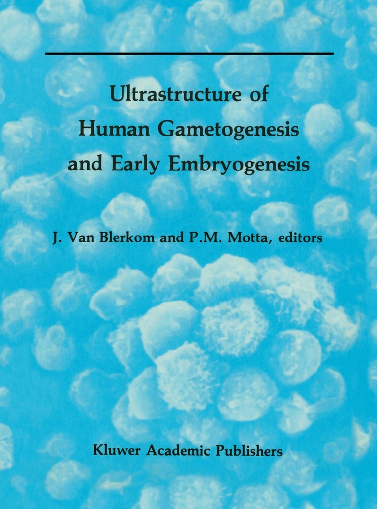
In the last few years, the adoption and worldwide proliferation of clinical procedures for medically assisted conception have been associated with the examination and analysis of spermatozoa, oocytes and early embryos under a variety of in vivo and in vitro conditions. These analyses have enabled correlations to be made between the behavior of gametes, the pattern of early embryonic development and the initiation of a normal pregnancy. Collectively, the findings have not only enormously increased our understanding of the process of early human development, but also have provided new insights into the origin and causes of reproductive failure in man. The research presented in this volume describes recent results derived from the study of normal and abnormal patterns of human spermatogenesis, oogenesis and early embryogenesis. The chapters discuss aberrations in morphodynamic and morphophysiological processes that have clinical relevance in human infertility and conception. Two of the chapters describe, respectively, the basic research that allows the cryopreservation of human oocytes and embryos, and the development of in vitro systems that permit the study of cell differentiation and interaction during the peri-implantation period. When relevant, each chapter extrapolates findings from in vitro experimentation to the comparable situation that is observed in vivo.
Inbunden, Engelska, 1988
2 223 kr
Skickas inom 10-15 vardagar
When established four years ago, the scope of this international series in electron microscopy essentially was to provide an opportunity for the pUblication of selected review contributions by specialists in ultrastructural research. Previous volumes presented over the last three years have focused on special topics of present interest in ~'ontemporary biomedicine such as endocrine cells, reproduction, and connective tissues. In these fielCls, in fact, integrated methods of electron microscopy have contributed much to generate new ideas and concepts of general value in both basic and clinical applications. The Ultrastructure of the Digestive Tract basically follows the same guidelines and style of the other books in the series and is an invited collection of selected contributions of authors from various laboratories active in the field of electron microscopy. Therefore, although the various chapters consist of individual topics, they nevertheless should be considered as interrelated contributions of specific subjects in the field. The idea was to have critical reviews of aspects previously published elsewhere by experts in the field who, as a rule, include other relevant information in their articles in order to update and enrich the subject. This book contains fifteen chapters by renowned electron microscopists. Each chapter, according to the policy of the editors, reviews a particular topic in great detail, providing updated information, study methods and results, authors' ideas on future investigative approaches, and possible guidelines for forthcoming work. We hope that this book will be useful to cell biologists, morphologists, physiologists, and pathologists.
Häftad, Engelska, 2011
1 115 kr
Skickas inom 10-15 vardagar
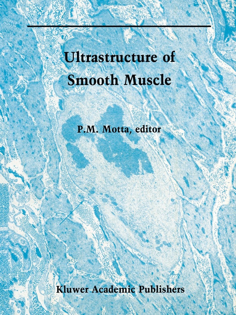
Recent advances in electron microscopy have opened up new dimensions and perspectives in the field of morphology, and these are presently being integrated with biochemical and physiopathological phenomena occurring in cells, tissues, and organs. Methods such as freeze-fracture, freeze-etching, scanning, and high-voltage electron microscopy have contributed immensely to this progress, as well as to the study of smooth muscle tissue and contractile cells in general. The articles composing this book have been selected and edited with the purpose of updating and reviewing the most important aspects of smooth muscle cells as revealed by the integration of these submicroscopic techniques. The chapters of this volume have been prepared by some of the most authoritative experts in the discipline. Therefore each article not only offers the reader a concise review of the specific topic, but also seeks to highlight areas that require further investigation. Much of the volume is presented in an illustrative format so as to emphasize the remarkable results obtainable by the combination of the aforementioned methods, which allow a better appreciation of smooth muscle structure and ultrastructure. This volume, like others in the series, is intended not only for researchers in the field, but also for graduate students of histology, embryology, anatomy, physiology, and pathology in both medical and veterinary colleges. My hope is that this book will prove to be a valuable academic resource to the audience of the world in this fascinating and expanding field.
Häftad, Engelska, 2012
1 115 kr
Skickas inom 10-15 vardagar
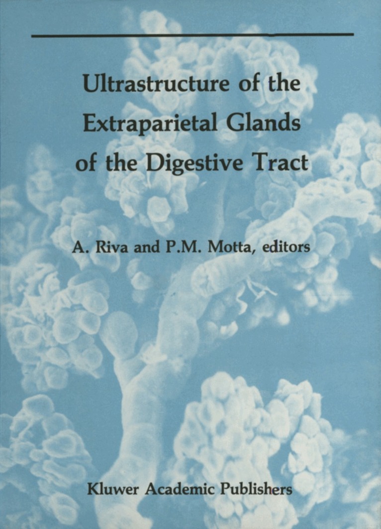
This volume, the sixth of the series, represents the natural counterpart of the previous volume, Ultra structure of the Digestive Tract. Unlike the latter, however, whose contents fell entirely within the domains of gastroenterology, Ultrastructure of the Extraparietal Glands of the Digestive Tract encom passes a few cognate sciences, such as hepatology, pancreatology, and even oral biology, which are usually dealt with separately. This allows, starting from cell biology, embryology, and comparative anatomy, a comprehensive survey of organs that have much in common both structurally and functionally. The chapters of this book have been compiled by well-known experts in the field with the aim not only of reviewing and pointing out the state of the art of the subject covered, but also of giving directions for future work. Furthermore, through the integration of electron microscopy with immunocytochemistry, autoradiography, freeze fracture, maceration, enzymatic digestion, etc., and by providing superb illus trative material, the authors substantiate the pivotal role played by modern morphology in under standing human physiology and pathology. In fact, it must be stressed, that a consistent part of the tissues studied here are from human origin. We believe that this volume should be read, not only by scientists and teachers active in the field, but also by a larger audience of students and professionals interested in knowing the scientific foundations of biomedicine.
Häftad, Engelska, 2011
1 669 kr
Skickas inom 10-15 vardagar
In the last few years, the adoption and worldwide proliferation of clinical procedures for medically assisted conception have been associated with the examination and analysis of spermatozoa, oocytes and early embryos under a variety of in vivo and in vitro conditions. These analyses have enabled correlations to be made between the behavior of gametes, the pattern of early embryonic development and the initiation of a normal pregnancy. Collectively, the findings have not only enormously increased our understanding of the process of early human development, but also have provided new insights into the origin and causes of reproductive failure in man. The research presented in this volume describes recent results derived from the study of normal and abnormal patterns of human spermatogenesis, oogenesis and early embryogenesis. The chapters discuss aberrations in morphodynamic and morphophysiological processes that have clinical relevance in human infertility and conception. Two of the chapters describe, respectively, the basic research that allows the cryopreservation of human oocytes and embryos, and the development of in vitro systems that permit the study of cell differentiation and interaction during the peri-implantation period. When relevant, each chapter extrapolates findings from in vitro experimentation to the comparable situation that is observed in vivo.
Häftad, Engelska, 2011
561 kr
Skickas inom 10-15 vardagar
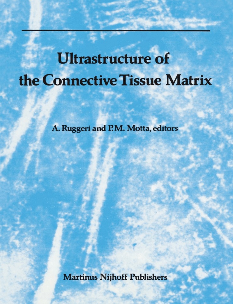
In recent years, the techniques of electron microscopy have developed so widely and rapidly that they now cover the fields of research once the unique ll:panage of sister research techniques such as biochemistry, physiology, immunology, X-ray diffraction, etc. It is now possible to reach molecular and submolecular levels, making this technique indispensable in every type of research. Electron microscopy alone often provides enough information to solve given problems. In the field of the connective tissue matrix, knowledge of the molecular structure of collagen, pro teoglycans and elastin and their interaction has been to a large extent elucidated by electron microscopy. The field over which electron microscopy ranges in the investigation of the connective tissue matrix is so wide that the aim of this volume is to collect the main ultrastructural acquisitions disseminated in various journals and monographs in one book. The intent ofthis volume is to: (a) integrate different and new microscopic methods and review the results of such an integrative approach; (b) present a comprehensive ultrastructural account of selected aspects of the field; (c) point out gaps or controversial topics in our knowledge; (d) outline pertinent future research and expansion of the subject.
Häftad, Engelska, 2011
561 kr
Skickas inom 10-15 vardagar
Innovative microscopic techniques, introduced during the last two decades, have contributed much to creating a new picture of the dynamic architecture of the cell, which can now be more exactly correlated with specific biochemical and physiopathological events. These developments have led to significant advances in our understanding of the physiomorphological and pathological aspects of the secretory mechanism, as well as the pharmacologic methods used to control, experimentally, the function of exocrine and endocrine glands. The integration of new ultrastructural methods such as freeze-fracture/etching, immunocytochemistry, scanning and high-voltage electron microscopy, cytoautoradiography, etc. , has proven to be of great value when applied to the study of endocrine cells and tissues. Because information on this topic has appeared in a variety of scientific and medical journals, this book: (1) reviews the results of an integrative approach presenting a comprehensive ultrastructural account of the main aspects of the field; (2) points out gaps or controversial topics in our knowledge; and (3) outlines pertinent directions for future research. The chapters, prepared by recognized authorities in the field, present traditional information on the topic in a concise manner and, with a valuable selection of original illustrations, show what the integration of new microscopic methods can contribute to the subject in terms of new concepts. This volume will be useful to cell biologists, anatomists, embryologists, histologists, pharmacologists, pathologists, and, of course, endocrinologists. It will also be of interest to students, practitioners of medicine, and to all others dealing with clinical research and diagnosis.
Del 2 - Electron Microscopy in Biology and Medicine
Ultrastructure of Reproduction
Gametogenesis, Fertilization, and Embryogenesis
Häftad, Engelska, 2011
561 kr
Skickas inom 10-15 vardagar
Advances in the development and application of electron microscopic techniques have occurred recently such that the electron microscope has evolved to become an essential tool in both basic and clinical research. Use of this instrument has contributed significantly to the formation of new perspectives and concepts concerning cell fine structure. These structural perspectives are now being integrated with specific functional, biochemical and pathophysiological events and processes of cells and tissues. Most recently, utilization of innovative electron microscopic techniques such as freeze-fracture, freeze etching, and scanning and high-voltage electron microscopy offers both the basic and clinical scientist potentially fundamental insights into many morphodynamic processes related to the activities of cells and tissues. Such an approach has been especially rewarding when applied to the dynamic events of gametogenesis and early embryonic development. The chapters comprising this book have been selected and edited with the aim of providing an up-to-date and comprehensive account of the most important aspects of vertebrate gamets and embryos as revealed by the integration of several different submicroscopic methods. The organization of the chapters is designed to indicate present gaps in our knowledge of the developmental and reproductive biology of gametes and the developing embryo and possible Iines of research which may lead to a lessening of these gaps.
Del 11 - Electron Microscopy in Biology and Medicine
Ultrastructure of the Male Urogenital Glands
Prostate, Seminal Vesicles, Urethral, and Bulbourethral Glands
Häftad, Engelska, 2012
561 kr
Skickas inom 10-15 vardagar
Male urogenital glands (also named male accessory sex glands) have received relatively little attention from electron microscopists, with the possible exception of the prostate gland. Moreover, even though comparative studies have clearly shown that these glands exhibit species-dependent features, very few studies, scattered over various publications, are available on the urogenital glands of man. This volume, the 11th of the series on Electron Microscopy in Biology and Medicine, presents an unprecedented collection of information on the functional microanatomy and cytoarchitecture of these organs in humans. Through the integration of transmission and scanning electron microscopy with a variety of modern techniques, it documents the most important aspects of the histophysiology of these glands from their development to some pathological alterations. In order to cover some key mechanisms of their cell biology, such as the action of sex hormones, the epithelio-mesenchymal interactions, and the dynamic of the secretory process, reports on human organs have been supplemented by some studies on experimental animals. The outstanding level of the contributions and the quality of the illustrations make this book, which has been compiled by some of the world authorities on the topic, a work of reference for students, scientists, and professionals interested in biomedical foundations of andrology, as well as a stimulus for future research in this exciting and relatively neglected chapter of human reproduction.
Häftad, Engelska, 2012
561 kr
Skickas inom 10-15 vardagar
Recently, attention has been called to the role that microvascular organization plays in the functional morphology of all organs and tissues, both in normal and pathological conditions. Since its development by Murakami, the corrosion cast method for scanning electron microscopy has come to be considered one of the most efficient means in clarifying the three-dimensional features of the microcirculation of organs and tissues. Scanning Electron Microscopy of Vascular Casts: Methods and Applications was planned to supply fundamental and new information regarding microcirculation studies to general biologists, anatomists, pathologists and clinicians. The contributions to this volume, contain original findings and excellent electron micrographs obtained by using recently improved corrosion cast methods. The rich variety of papers in this book will be useful to many, and will provide both the basic and clinically oriented readers with good ideas, suggestions, and original and worthwhile information.

Häftad, Engelska, 2012
3 331 kr
Skickas inom 10-15 vardagar

Del 1 - Electron Microscopy in Biology and Medicine
Ultrastructure of Endocrine Cells and Tissues
Häftad, Engelska, 1984
545 kr
Skickas inom 10-15 vardagar
Innovative microscopic techniques, introduced during the last two decades, have contributed much to creating a new picture of the dynamic architecture of the cell, which can now be more exactly correlated with specific biochemical and physiopathological events. These developments have led to significant advances in our understanding ofthe physiomorphological and pathological aspects of the secretory mechanism, as well as the pharmacologic methods used to control, experimentally, the function of exocrine and endocrine glands. The integration of new ultrastructural methods such as freeze-fracturejetching, immunocytochemistry, scanning and high-voltage electron microscopy, cytoautoradiography, etc. , has proven to be of great value when applied to the study of endocrine cells and tissues. Because information on this topic has appeared in a variety of scientific and medical journals, this book: (1) reviews the results of an integrative approach presenting a comprehensive ultrastructural account of the main aspects of the field; (2) points out gaps or controversial topics in our knowledge; and (3) outlines pertinent directions for future research. The chapters, prepared by recognized authorities in the field, present traditional information on the topic in a concise manner and, with a valuable selection of original illustrations, show what the integration of new microscopic methods can contribute to the subject in terms of new concepts. This volume will be useful to cell biologists, anatomists, embryologists, histologists, pharmacologists, pathologists, and, of course, endocrinologists. It will also be of interest to students, practitioners of medicine, and to all others dealing with clinical research and diagnosis.