
A. L. Baert – författare
Visar alla böcker från författaren A. L. Baert. Handla med fri frakt och snabb leverans.
9 produkter
9 produkter
E-bok
PDF, Engelska, 20121 132 kr
Läs direkt efter köp
It was our aim to place at the disposal of radiologists within a short time an atlas of high-quality, valuable pictures of abdominal CT without We felt that, the image degradation inherent to slower scanning apparatus. notwithstanding rapid evolution in CT scanning apparatus and the resulting rapid advances in our clinical knowledge about the value, limitations and applications of this new diagnostic imaging modality, an effort should be made to realize an atlas of reference. From more than 7,000 patients studied with abdominal CT, we tried to assemble images with maximal anatomical detail, which implies the use of large window settings, being well aware that in daily routine practice basic CT methodology includes the use of different window settings for optimal information. It was, however, a conscious decision not to use comparative images with large and small window settings in order to stay within a reasonable total number of figures. Much emphasis has been placed upon the use of contrast enhancement by intravenous contrast media. This is based on the conviction that essen tially new and better morphological information about normal and pathological processes within the abdomen can be obtained because short exposure times now allow one to capture the rapidly changing aspect and degree of contrast enhancement of the lesions as a function of their vascu larity.
E-bok
PDF, Engelska, 20121 367 kr
Läs direkt efter köp
Häftad, Engelska, 2012
1 092 kr
Skickas inom 10-15 vardagar
With contributions by numerous experts
E-bok
PDF, Engelska, 20121 420 kr
Läs direkt efter köp
Häftad, Engelska, 2011
1 125 kr
Skickas inom 10-15 vardagar
E-bok
PDF, Engelska, 20121 408 kr
Läs direkt efter köp
Häftad, Engelska, 2011
1 125 kr
Skickas inom 10-15 vardagar
Contents Abdominal and Thoracic Magnetic Resonance Angiography: Challenges, Technical Approaches and First Clinical Applications. H.U. Kauczor, R. Hausmann, W. Semmler Magnetic Resonance Spectroscopy of Skeletal Muscle. F. Tr{ber, W.A. Kaiser, G. Layer,C. Kuhl, M. Reiser Hepatic Tumors: Relaxometry and Quantitative Tissue Characterization with Magnetic Resonance Imaging. A. Steudel, F. Tr{ber, M. Reiser Liver-Specific Particulate Contrast Agents: An Overview I. Ivancev, A. Lunderquist Contrast Medium Induced Nephropathy:Animal Experiments H.S. Thomsen et al. Rheological Properties of Blood after Diagnostic Cardiac Catheterisation with Iopromide. R. Bach et al. Color Coded Doppler Sonography of the Male Urethra During Voiding: New Observations of Flow in an Aqueous Medium and Their Physical Foundations. P. H}bsch, F. Hager, E. Benes Development of a Model of Arterial Stenosis Analysis Using Digital Subtraction Angiography: A Parametric Study of Image Quality. C. Renaudin, I. Magnin, A. Amiel Arterial Calcifications in Thoracoabdominal Computer Tomography: Frequency and Correlation with Risk Factors and Cardiovascular Diseases. J. G|rich et al.

E-bok
PDF, Engelska, 2013708 kr
Läs direkt efter köp
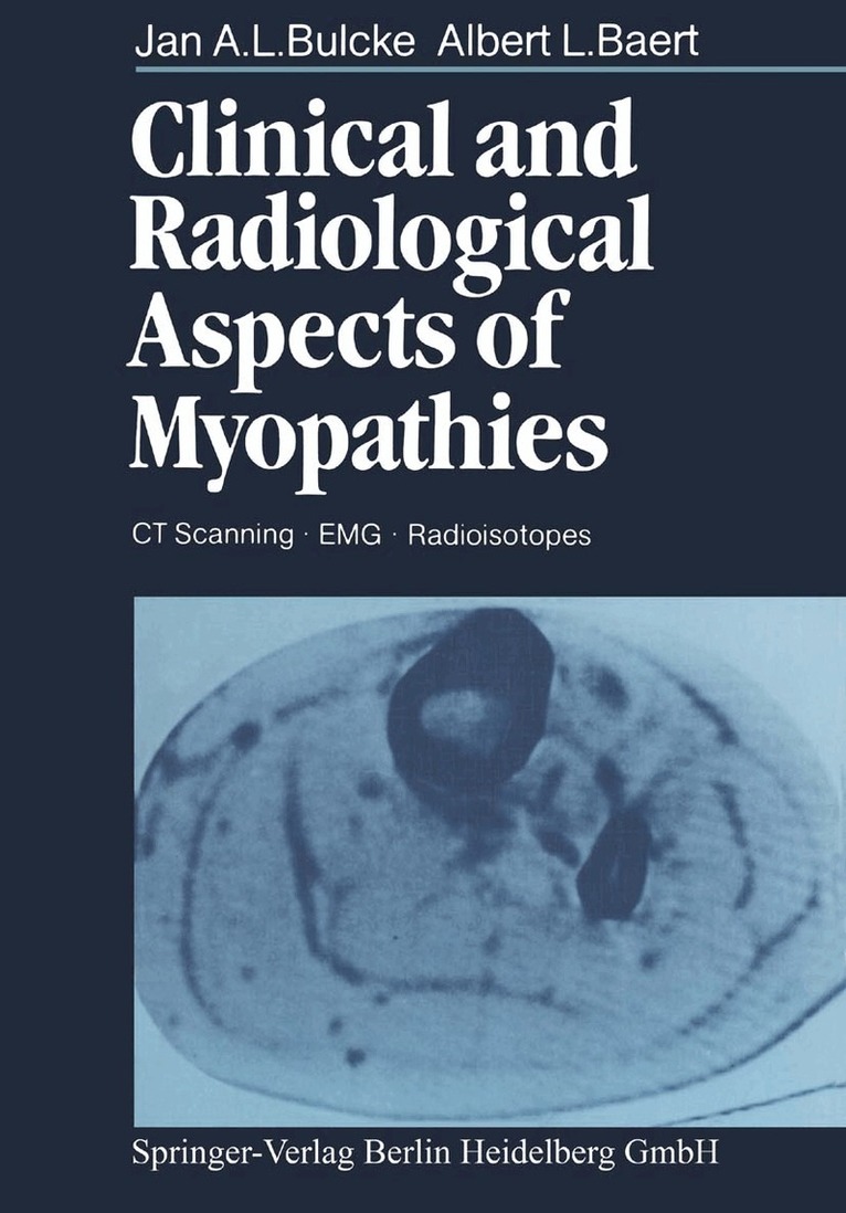
One of the most puzzling and striking features of many of the genetically determined progressive neuromuscular diseases such as the spinal muscular atrophies and the muscular dystrophies is that muscular wasting and weak ness in these cases is curiously selective, at least in the early stages, pick ing out certain skeletal muscles and sparing others. The diagnosis of these conditions has largely depended in the past upon the recognition of specific patterns of involvement of individual muscles and muscle groups, taken along with information derived from the mode of inheritance within the in dividual family and the results of special investigations. The investigations of most value have proved to be serum enzyme studies, electromyography and related techniques, and muscle biopsy. The advent of CT scanning has, however, introduced a new dimension; as the authors of this interesting monograph have clearly demonstrated, it is now possible, using the whole body scanner, to define patterns of muscular atrophy in the limbs and trunk much more precisely than by any other method. Not only does this techni que demonstrate which muscles are involved, but the changes in relative density provide useful information about the severity of the process and about the progress of the disease if the studies are performed serially. This monograph is pleasantly written and most attractively illustrated.
Häftad, Engelska, 2013
565 kr
Skickas inom 10-15 vardagar
One of the most puzzling and striking features of many of the genetically determined progressive neuromuscular diseases such as the spinal muscular atrophies and the muscular dystrophies is that muscular wasting and weak ness in these cases is curiously selective, at least in the early stages, pick ing out certain skeletal muscles and sparing others. The diagnosis of these conditions has largely depended in the past upon the recognition of specific patterns of involvement of individual muscles and muscle groups, taken along with information derived from the mode of inheritance within the in dividual family and the results of special investigations. The investigations of most value have proved to be serum enzyme studies, electromyography and related techniques, and muscle biopsy. The advent of CT scanning has, however, introduced a new dimension; as the authors of this interesting monograph have clearly demonstrated, it is now possible, using the whole body scanner, to define patterns of muscular atrophy in the limbs and trunk much more precisely than by any other method. Not only does this techni que demonstrate which muscles are involved, but the changes in relative density provide useful information about the severity of the process and about the progress of the disease if the studies are performed serially. This monograph is pleasantly written and most attractively illustrated.