
Ammasi Periasamy – författare
Visar alla böcker från författaren . Handla med fri frakt och snabb leverans.
7 produkter
7 produkter
E-bok
PDF, Engelska, 20111 203 kr
Läs direkt efter köp
The detection and measurement of the dynamic interactions of proteins within the living cell are critical to our understanding of cell physiology and pathophysiology. With FRET microscopy and spectroscopy techniques, basic and clinical scientists can make such measurements at very high spatial and temporal resolution. But sources of background information about these tools are very limited, so this book fills an important gap. It covers both the basic concepts and theory behind the various FRET microscopy and spectroscopy techniques, and the practical aspects of using the techniques and analyzing the results. The critical tricks for obtaining a good FRET image and precisely quantitating the signals from living specimens at the nanomolecular level are explained. Valuable information about the preparation of biological samples used for FRET image analysis is also provided. The methods covered include different types of microscopy systems and detectors (wide-field, confocal, multi-photon) as well as specialized techniques such as photobleaching FRET, FLIM-FRET microscopy, spectral imaging FRET, single molecule FRET, and time and image correlation spectroscopy. Starting from the basics, the chapters guide readers through the choice of probes to be used for FRET experiments and the selection of the most suitable experimental approaches to address specific biological questions. Up-to-date, consistently organized, and well-illustrated, this unique book will be welcomed by all researchers who wish to use FRET microscopy and spectroscopy techniques.
Inbunden, Engelska, 2008
1 329 kr
Skickas inom 10-15 vardagar
The detection and measurement of the dynamic interactions of proteins within the living cell are critical to our understanding of cell physiology and pathophysiology. With FRET microscopy and spectroscopy techniques, basic and clinical scientists can make such measurements at very high spatial and temporal resolution. But sources of background information about these tools are very limited, so this book fills an important gap. It covers both the basic concepts and theory behind the various FRET microscopy and spectroscopy techniques, and the practical aspects of using the techniques and analyzing the results. The critical tricks for obtaining a good FRET image and precisely quantitating the signals from living specimens at the nanomolecular level are explained. Valuable information about the preparation of biological samples used for FRET image analysis is also provided. The methods covered include different types of microscopy systems and detectors (wide-field, confocal, multi-photon) as well as specialized techniques such as photobleaching FRET, FLIM-FRET microscopy, spectral imaging FRET, single molecule FRET, and time and image correlation spectroscopy. Starting from the basics, the chapters guide readers through the choice of probes to be used for FRET experiments and the selection of the most suitable experimental approaches to address specific biological questions. Up-to-date, consistently organized, and well-illustrated, this unique book will be welcomed by all researchers who wish to use FRET microscopy and spectroscopy techniques.
Häftad, Engelska, 2020
765 kr
Skickas inom 10-15 vardagar
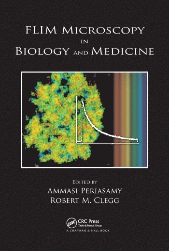
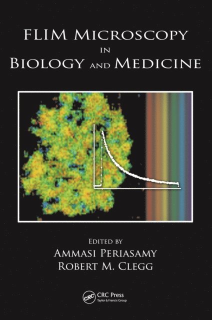
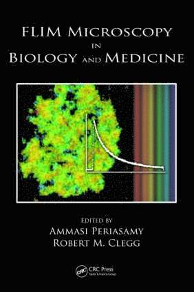
Detecting Signals at the Single Molecule Level: Pioneering Achievements in MicroscopyRecent advances have led to such remarkable improvements in fluorescence lifetime imaging microscopy’s (FLIM) capacity for contrast and sensitivity that researchers can now employ it to detect signals at the single molecule level. FLIM also offers the additional benefit of independence from fluorophore concentration and excitation intensity. Moreover, its unique sensitivity makes it an excellent reporter of conformational changes and of variations in the molecular surroundings of biological molecules. Most of this improvement and discovery have occurred during the past decade, and, to date, information that would benefit a broad range of researchers remains scattered in the literature. Edited by two of the top pioneers in the field, FLIM Microscopy in Biology and Medicine presents the fundamentals of FLIM along with a number of advanced considerations so that a wider audience can appreciate recent and potential improvements that make it such a valuable tool. New Opportunities for Biomedical Researchers… New Challenges for Microscopy ResearchersDiscussion sections in all the chapters clearly show the challenges for implementing FLIM for various applications. Certain chapters discuss limits on the number of photons required for highly accurate lifetime determinations, as well as the accuracy with which multiple, closely associated lifetime components can reliably be determined. Such considerations are important for the user when he or she is selecting the most advantageous method of FLIM to use for a particular application. While this book provides an introduction for those new to FLIM, it gathers a wealth of material to enhance the work of experts involved in pioneering technological improvements, as well as those research opportunities in this unique and promising area of microscopy.
E-bok
Engelska, 2009935 kr
Läs direkt efter köp
Detecting Signals at the Single Molecule Level: Pioneering Achievements in MicroscopyRecent advances have led to such remarkable improvements in fluorescence lifetime imaging microscopy''s (FLIM) capacity for contrast and sensitivity that researchers can now employ it to detect signals at the single molecule level. FLIM also offers the additional be
Inbunden, Engelska, 2009
2 955 kr
Skickas inom 10-15 vardagar
Detecting Signals at the Single Molecule Level: Pioneering Achievements in MicroscopyRecent advances have led to such remarkable improvements in fluorescence lifetime imaging microscopy’s (FLIM) capacity for contrast and sensitivity that researchers can now employ it to detect signals at the single molecule level. FLIM also offers the additional benefit of independence from fluorophore concentration and excitation intensity. Moreover, its unique sensitivity makes it an excellent reporter of conformational changes and of variations in the molecular surroundings of biological molecules. Most of this improvement and discovery have occurred during the past decade, and, to date, information that would benefit a broad range of researchers remains scattered in the literature. Edited by two of the top pioneers in the field, FLIM Microscopy in Biology and Medicine presents the fundamentals of FLIM along with a number of advanced considerations so that a wider audience can appreciate recent and potential improvements that make it such a valuable tool. New Opportunities for Biomedical Researchers… New Challenges for Microscopy ResearchersDiscussion sections in all the chapters clearly show the challenges for implementing FLIM for various applications. Certain chapters discuss limits on the number of photons required for highly accurate lifetime determinations, as well as the accuracy with which multiple, closely associated lifetime components can reliably be determined. Such considerations are important for the user when he or she is selecting the most advantageous method of FLIM to use for a particular application. While this book provides an introduction for those new to FLIM, it gathers a wealth of material to enhance the work of experts involved in pioneering technological improvements, as well as those research opportunities in this unique and promising area of microscopy.

E-bok
PDF, Engelska, 2009935 kr
Läs direkt efter köp
Detecting Signals at the Single Molecule Level: Pioneering Achievements in MicroscopyRecent advances have led to such remarkable improvements in fluorescence lifetime imaging microscopy''s (FLIM) capacity for contrast and sensitivity that researchers can now employ it to detect signals at the single molecule level. FLIM also offers the additional be
E-bok
PDF, Engelska, 20131 258 kr
Läs direkt efter köp
Advances in technology have revolutionized the development of light microscopy techniques in biomedical research, thus improving visualization of the microstructure of cells and tissues under physiological conditions. Fluorescence microscopy methods are non-contact and non-invasive and provide high spatial and temporal resolution that other laboratory techniques cannot. This well-illustrated book targets graduate students and scientists who are new to the state-of-the-art fluorescence microscopy techniques used in biological and clinical imaging. It explains basic concepts and imaging procedures for wide-field, confocal, multiphoton excitation, fluorescence resonance energy transfer (FRET), lifetime imaging (FLIM), spectral imaging, fluorescence recovery after photobleaching (FRAP), optical tweezers, total internal reflection, high spatial resolution atomic force microscopy (AFM), and bioluminescence imaging for gene expression. The usage of these techniques in various biological applications, including calcium, pH, membrane potential, mitochondrial signaling, protein-protein interactions under various physiological conditions, and deep tissue imaging, is clearly presented. The authors describe the approaches to selecting epifluorescence microscopy, the detectors, and the image acquisition and processing software for different biological applications. Step-by-step directions on preparing different digital formats for light microscopy images on websites are also provided.