
Atif Zaheer – författare
Visar alla böcker från författaren Atif Zaheer. Handla med fri frakt och snabb leverans.
7 produkter
7 produkter
E-bok
Engelska, 20224 138 kr
Läs direkt efter köp
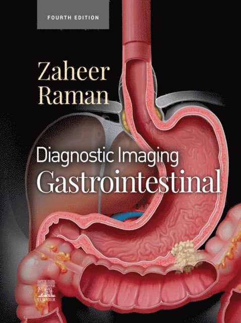
Covering the entire spectrum of this fast-changing field, Diagnostic Imaging: Gastrointestinal, fourth edition, is an invaluable resource for gastrointestinal radiologists, general radiologists, and trainees—anyone who requires an easily accessible, highly visual reference on today''s GI imaging. Drs. Siva P. Raman, Atif Zaheer, and their team of highly regarded experts provide up-to-date information on recent advances in technology and the understanding of GI diseases and disorders to help you make informed decisions at the point of care. The text is lavishly illustrated, delineated, and referenced, making it a useful learning tool as well as a handy reference for daily practice.- Serves as a one-stop resource for key concepts and information on gastrointestinal imaging, including a wealth of new material and content updates throughout- Features more than 2,900 illustrations (multiplanar CT, sonography, MR, and PET/CT; clinical photos; radiologic images; histologic images; H&E stains; and full-color illustrations) as well as an additional 3K digital-only images and new video clips- Features updates from cover to cover including new information on MRI imaging of rectal cancer, iron quantification, and MRI protocols; new cases and images, and new staging details and diagrams- Contains new chapters on treatment response criteria for systemic conditions (RECIST, irRECIST, etc.), dual energy CT for pancreas, vascular abnormalities, MR elastography of the liver, pretransplant liver evaluation, and more- Covers all aspects of GI imaging, including pathophysiology, imaging findings, and disease management options such as the radiologist''s role in evaluating patients for bariatric surgery, antireflux procedures, esophageal and bowel resections, and more- Uses bulleted, succinct text and highly templated chapters for quick comprehension of essential information at the point of care
Inbunden, Engelska, 2022
2 920 kr
Skickas inom 5-8 vardagar
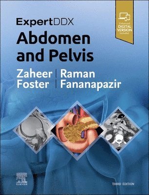
Highly practical and user-friendly,�ExpertDDx: Abdomen and Pelvis,�third edition, helps you�reach accurate, clinically useful differential diagnoses�in your everyday practice. It presents the most useful differential diagnoses for each region of the abdomen and pelvis, grouped according to anatomic location, generic imaging findings, modality-specific findings, or clinical-based indications. Each differential diagnosis includes several�high-quality, succinctly annotated images; a�list of diagnostic possibilities�sorted as common, less common, and rare but important; and�brief, bulleted text�offering helpful diagnostic clues. It's an excellent resource for subspecialty abdominal imagers as well as general radiologists and trainees, providing invaluable assistance in reaching logical, on-target differential diagnoses based on key imaging findings and clinical details.
Inbunden, Engelska, 2023
3 433 kr
Skickas inom 5-8 vardagar
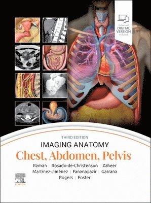
This richly illustrated and superbly organized text/atlas is an excellent point-of-care resource for practitioners at all levels of experience and training. Written by global leaders in the field, Imaging Anatomy: Chest, Abdomen, Pelvis, third edition, contains specifics about radiographic, multiplanar, high-resolution, and cross-sectional body imaging along with thousands of relevant examples to give busy clinicians quick answers to imaging anatomy questions. This must-have reference employs a templated, highly formatted design; concise, bulleted text; and state-of-the-art images throughout that identify characteristic normal imaging findings and anatomic variants in each anatomic area, offering a unique opportunity to master the fundamentals of normal anatomy and accurately and efficiently recognize pathologic conditions.
Inbunden, Engelska, 2025
3 715 kr
Skickas inom 5-8 vardagar
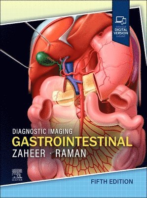
Covering the entire spectrum of this fast-changing field, the fifth edition of Diagnostic Imaging: Gastrointestinal is an invaluable resource for GI radiologists, general radiologists, and trainees?anyone who requires an easily accessible, highly visual reference on today’s GI imaging. Drs. Atif Zaheer, Siva P. Raman, and their team of highly regarded experts provide updated information on recent advances in technology and the understanding of GI diseases and disorders to help you make informed decisions at the point of care. The text is image-rich, with succinct bullets that quickly convey details, and includes the latest literature references, making it a useful learning tool as well as a handy reference for daily practice.
Inbunden, Engelska, 2017
2 437 kr
Skickas inom 10-15 vardagar
This comprehensive teaching atlas covers virtually all pancreatic anatomy (including variants) and diseases in a pattern-based radiologic approach. Cases are presented as “unknowns”, allowing the reader to analyze the findings and learn key points. Each teaching case includes a brief clinical history, images, a description of imaging findings, differential diagnoses, final diagnosis with images of gross pathology, and a discussion of key teaching points. The presented images have been acquired with the full range of relevant modalities, including state of the art technologies such as multidetector row dual-phase CT, 3D reformatting, and multiple MRI sequences. The book will help radiologists, radiology residents and fellows to sharpen their diagnostic skills by looking at a vast array of pathology from a major tertiary hospital (Johns Hopkins) and will also assist in preparation for radiology board examinations.
E-bok
Engelska, 20172 273 kr
Läs direkt efter köp
This comprehensive teaching atlas covers virtually all pancreatic anatomy (including variants) and diseases in a pattern-based radiologic approach. Cases are presented as “unknowns”, allowing the reader to analyze the findings and learn key points. Each teaching case includes a brief clinical history, images, a description of imaging findings, differential diagnoses, final diagnosis with images of gross pathology, and a discussion of key teaching points. The presented images have been acquired with the full range of relevant modalities, including state of the art technologies such as multidetector row dual-phase CT, 3D reformatting, and multiple MRI sequences. The book will help radiologists, radiology residents and fellows to sharpen their diagnostic skills by looking at a vast array of pathology from a major tertiary hospital (Johns Hopkins) and will also assist in preparation for radiology board examinations.
Häftad, Engelska, 2018
1 995 kr
Skickas inom 10-15 vardagar
This comprehensive teaching atlas covers virtually all pancreatic anatomy (including variants) and diseases in a pattern-based radiologic approach. Cases are presented as “unknowns”, allowing the reader to analyze the findings and learn key points. Each teaching case includes a brief clinical history, images, a description of imaging findings, differential diagnoses, final diagnosis with images of gross pathology, and a discussion of key teaching points. The presented images have been acquired with the full range of relevant modalities, including state of the art technologies such as multidetector row dual-phase CT, 3D reformatting, and multiple MRI sequences. The book will help radiologists, radiology residents and fellows to sharpen their diagnostic skills by looking at a vast array of pathology from a major tertiary hospital (Johns Hopkins) and will also assist in preparation for radiology board examinations.