Elliot K. Fishman – författare
Visar alla böcker från författaren Elliot K. Fishman. Handla med fri frakt och snabb leverans.
6 produkter
6 produkter
Pearls and Pitfalls in Cardiovascular Imaging
Pseudolesions, Artifacts, and Other Difficult Diagnoses
E-bok
Engelska, 20151 858 kr
Läs direkt efter köp
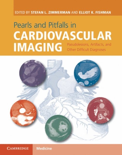
Cardiovascular imagers are faced with the challenge of interpreting cases that include artifacts, unusual findings, or anatomic variants on an almost daily basis. These studies can result in confusion and may lead to misdiagnosis even for the most experienced imager. This book provides an approachable reference for practising cardiovascular imagers to aid with both commonly and uncommonly encountered entities that can result in inappropriate patient management. Through the focused use of case examples, this book reviews 100 conditions that can be seen in clinical practice, including pseudotumors, artifacts, anatomic variants, mimics, and unusual diagnoses. Each highly illustrated case follows a standard format, allowing readers to learn from real-life examples and provides an accessible and rapid source of reference for the improved interpretation of cardiovascular imaging and enhanced patient care. This text will be invaluable to radiologists, cardiologists, and trainees.
Pearls and Pitfalls in Cardiovascular Imaging
Pseudolesions, Artifacts, and Other Difficult Diagnoses
E-bok
PDF, Engelska, 20151 793 kr
Läs direkt efter köp
Cardiovascular imagers are faced with the challenge of interpreting cases that include artifacts, unusual findings, or anatomic variants on an almost daily basis. These studies can result in confusion and may lead to misdiagnosis even for the most experienced imager. This book provides an approachable reference for practising cardiovascular imagers to aid with both commonly and uncommonly encountered entities that can result in inappropriate patient management. Through the focused use of case examples, this book reviews 100 conditions that can be seen in clinical practice, including pseudotumors, artifacts, anatomic variants, mimics, and unusual diagnoses. Each highly illustrated case follows a standard format, allowing readers to learn from real-life examples and provides an accessible and rapid source of reference for the improved interpretation of cardiovascular imaging and enhanced patient care. This text will be invaluable to radiologists, cardiologists, and trainees.
Inbunden, Engelska, 2017
2 444 kr
Skickas inom 10-15 vardagar
This comprehensive teaching atlas covers virtually all pancreatic anatomy (including variants) and diseases in a pattern-based radiologic approach. Cases are presented as “unknowns”, allowing the reader to analyze the findings and learn key points. Each teaching case includes a brief clinical history, images, a description of imaging findings, differential diagnoses, final diagnosis with images of gross pathology, and a discussion of key teaching points. The presented images have been acquired with the full range of relevant modalities, including state of the art technologies such as multidetector row dual-phase CT, 3D reformatting, and multiple MRI sequences. The book will help radiologists, radiology residents and fellows to sharpen their diagnostic skills by looking at a vast array of pathology from a major tertiary hospital (Johns Hopkins) and will also assist in preparation for radiology board examinations.
E-bok
Engelska, 20172 273 kr
Läs direkt efter köp
This comprehensive teaching atlas covers virtually all pancreatic anatomy (including variants) and diseases in a pattern-based radiologic approach. Cases are presented as “unknowns”, allowing the reader to analyze the findings and learn key points. Each teaching case includes a brief clinical history, images, a description of imaging findings, differential diagnoses, final diagnosis with images of gross pathology, and a discussion of key teaching points. The presented images have been acquired with the full range of relevant modalities, including state of the art technologies such as multidetector row dual-phase CT, 3D reformatting, and multiple MRI sequences. The book will help radiologists, radiology residents and fellows to sharpen their diagnostic skills by looking at a vast array of pathology from a major tertiary hospital (Johns Hopkins) and will also assist in preparation for radiology board examinations.
Häftad, Engelska, 2018
2 001 kr
Skickas inom 10-15 vardagar
This comprehensive teaching atlas covers virtually all pancreatic anatomy (including variants) and diseases in a pattern-based radiologic approach. Cases are presented as “unknowns”, allowing the reader to analyze the findings and learn key points. Each teaching case includes a brief clinical history, images, a description of imaging findings, differential diagnoses, final diagnosis with images of gross pathology, and a discussion of key teaching points. The presented images have been acquired with the full range of relevant modalities, including state of the art technologies such as multidetector row dual-phase CT, 3D reformatting, and multiple MRI sequences. The book will help radiologists, radiology residents and fellows to sharpen their diagnostic skills by looking at a vast array of pathology from a major tertiary hospital (Johns Hopkins) and will also assist in preparation for radiology board examinations.
Inbunden, Engelska, 2003
893 kr
Skickas inom 10-15 vardagar
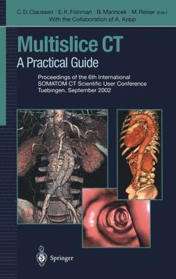
There have been remarkable achievements in CT technology, workflow management and applications in the last couple of years. The introduction of 4- and 16-row multidetector technology has substantially increased acquisition speed and provides nearly isotropic resolution. These new technical possibilities had significant impact on the clinical use of CT and have yielded a broadening of the spectrum of applications, particularly in vascular, cardiac, abdominal, and trauma imaging. This book presents the practical experience of an international expert group of radiologists and physicists with state-of-the-art multidetector technology. The chapters in this book will facilitate a thorough understanding of 4- and 16-slice multidetector-row CT and its clinical applications. This will help to fully exploit the diagnostic potential of this technology.