
Claus Claussen – författare
Visar alla böcker från författaren Claus Claussen. Handla med fri frakt och snabb leverans.
12 produkter
12 produkter

Häftad, Tyska
308 kr
Skickas inom 3-6 vardagar

361 kr
Skickas inom 3-6 vardagar

Häftad, Tyska, 1997
444 kr
Skickas inom 3-6 vardagar
Inbunden, Engelska, 2003
893 kr
Skickas inom 10-15 vardagar
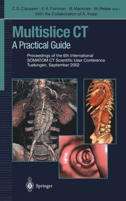
There have been remarkable achievements in CT technology, workflow management and applications in the last couple of years. The introduction of 4- and 16-row multidetector technology has substantially increased acquisition speed and provides nearly isotropic resolution. These new technical possibilities had significant impact on the clinical use of CT and have yielded a broadening of the spectrum of applications, particularly in vascular, cardiac, abdominal, and trauma imaging. This book presents the practical experience of an international expert group of radiologists and physicists with state-of-the-art multidetector technology. The chapters in this book will facilitate a thorough understanding of 4- and 16-slice multidetector-row CT and its clinical applications. This will help to fully exploit the diagnostic potential of this technology.
E-bok
PDF, Engelska, 20121 408 kr
Läs direkt efter köp
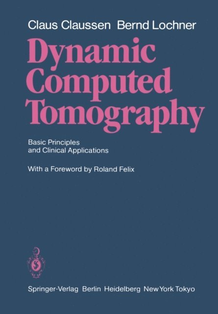
The authors present their experience in more than seven years of dynamic computed tomography in clinical practice. Time density curves and characteristic examples in specific regions of interest enrich the presentation. Dynamic computed tomography makes an important contribu tion to the diagnosis and evaluation of a pathologic process: the demonstration of the dynamics of blood flow within the lesion and surrounding normal tissue. Since both the lesion itself and adjacent normal tissue demonstrate characteristic findings in each circulatory phase, the study provides a large amount of data on the flow of blood and contrast material which facilitate both recognition and diiferentation of a lesion. Late studies following administration of a contrast agent allow an estimate of the passage of the contrast medium to the inter stitium, which is of diagnostic importance. Chapters dealing with specific clinical entities also contain useful information on the most appropriate means of contrast agent administration (bolus injection or infusion) as well as a discussion of indications for the procedure. Dynamic computed tomography represents a significant advance over conventional computed tomography in some situations, and this signifies a major contri bution to the diagnostic capabilities of the clinical radiologist. The authors are to be commended for the fact that they have clearly defined the limits of dynamic computed tomography. I hope that the first English language edition, following the appea rance of the German version in 1983, will be well received.

Häftad, Engelska, 2011
1 115 kr
Skickas inom 10-15 vardagar
The authors present their experience in more than seven years of dynamic computed tomography in clinical practice. Time density curves and characteristic examples in specific regions of interest enrich the presentation. Dynamic computed tomography makes an important contribu tion to the diagnosis and evaluation of a pathologic process: the demonstration of the dynamics of blood flow within the lesion and surrounding normal tissue. Since both the lesion itself and adjacent normal tissue demonstrate characteristic findings in each circulatory phase, the study provides a large amount of data on the flow of blood and contrast material which facilitate both recognition and diiferentation of a lesion. Late studies following administration of a contrast agent allow an estimate of the passage of the contrast medium to the inter stitium, which is of diagnostic importance. Chapters dealing with specific clinical entities also contain useful information on the most appropriate means of contrast agent administration (bolus injection or infusion) as well as a discussion of indications for the procedure. Dynamic computed tomography represents a significant advance over conventional computed tomography in some situations, and this signifies a major contri bution to the diagnostic capabilities of the clinical radiologist. The authors are to be commended for the fact that they have clearly defined the limits of dynamic computed tomography. I hope that the first English language edition, following the appea rance of the German version in 1983, will be well received.
E-bok
PDF, Tyska, 2013582 kr
Läs direkt efter köp
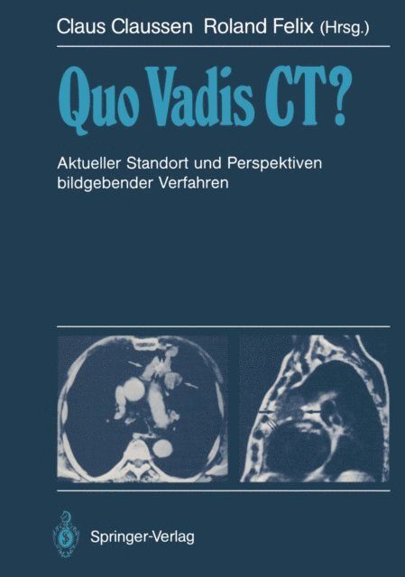
Die Computertomographie hat sich im letzten Jahrzehnt zu einem diagnostischen Routineverfahren entwickelt. Die rasche Entwicklung insbesondere der Kernspintomographie stellt die Frage: Wie geht es mit den bildgebenden Verfahren, vor allem mit der Röntgencomputertomographie, weiter? Bei vielen Radiologen und auch Ärzten anderer Disziplinen herrscht Unklarheit hinsichtlich des Leistungsspektrums der einzelnen bildgebenden Verfahren und deren zukünftigen Entwicklungen. Dieses Buch trägt dazu bei, die Möglichkeiten und Grenzen der Computertomographie beim heutigen Entwicklungsstand im Vergleich zu Ultraschall und Kernspintomographie darzulegen und die zukünftigen Perspektiven der CT im Vergleich zu den anderen Methoden aufzuzeigen.

Häftad, Tyska, 2011
576 kr
Skickas inom 10-15 vardagar
Die Computertomographie hat sich im letzten Jahrzehnt zu einem diagnostischen Routineverfahren entwickelt. Die rasche Entwicklung insbesondere der Kernspintomographie stellt die Frage: Wie geht es mit den bildgebenden Verfahren, vor allem mit der Röntgencomputertomographie, weiter? Bei vielen Radiologen und auch Ärzten anderer Disziplinen herrscht Unklarheit hinsichtlich des Leistungsspektrums der einzelnen bildgebenden Verfahren und deren zukünftigen Entwicklungen. Dieses Buch trägt dazu bei, die Möglichkeiten und Grenzen der Computertomographie beim heutigen Entwicklungsstand im Vergleich zu Ultraschall und Kernspintomographie darzulegen und die zukünftigen Perspektiven der CT im Vergleich zu den anderen Methoden aufzuzeigen.
E-bok
PDF, Engelska, 20121 459 kr
Läs direkt efter köp
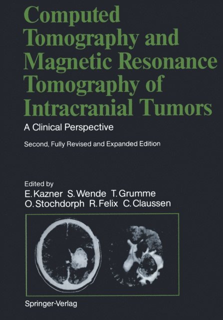
This book represents the second, fully revised edition of the original volume published in 1982. Experience in neuroradiology has confirmed the outstanding value of computed tomography (CT) for the diagnosis of space-occupying lesions within the skull and orbit. It might be assumed, then, that the second edition of this book would simply represent a numerically expanded continua tion of the popular first edition. That is not the case, however. Advances in imaging techniques have promp ted the creation of a new book whose expanded title reflects its more comprehen sive nature. The added illustrations, the revised text, and the expanded circle of editors and contributors document this. Since publication of the first edition, a new modality, magnetic resonance imaging (MRI), has become an established neuroradiologic study. We felt it was essential to include this new modality in our book and explore its capabilities as an adjunct or alternative to CT scanning. Because of the high acquisition costs of MRI and the still small number of MR units currently in operation, we have relied in part on images furnished by other institutions and private practitioners, to whom we are indebted. Many problems relating to MR, both in terms of equipment and image interpretation, have yet to be resolved. There is no denying that we still have much to learn.

Häftad, Engelska, 2011
1 115 kr
Skickas inom 10-15 vardagar
This book represents the second, fully revised edition of the original volume published in 1982. Experience in neuroradiology has confirmed the outstanding value of computed tomography (CT) for the diagnosis of space-occupying lesions within the skull and orbit. It might be assumed, then, that the second edition of this book would simply represent a numerically expanded continua tion of the popular first edition. That is not the case, however. Advances in imaging techniques have promp ted the creation of a new book whose expanded title reflects its more comprehen sive nature. The added illustrations, the revised text, and the expanded circle of editors and contributors document this. Since publication of the first edition, a new modality, magnetic resonance imaging (MRI), has become an established neuroradiologic study. We felt it was essential to include this new modality in our book and explore its capabilities as an adjunct or alternative to CT scanning. Because of the high acquisition costs of MRI and the still small number of MR units currently in operation, we have relied in part on images furnished by other institutions and private practitioners, to whom we are indebted. Many problems relating to MR, both in terms of equipment and image interpretation, have yet to be resolved. There is no denying that we still have much to learn.
E-bok
PDF, Tyska, 2013587 kr
Läs direkt efter köp
Dieses weltweit bewährte Standardwerk über die zerebrale Computertomographie wurde völlig neu überarbeitet und durch Einbeziehung der Kernspintomographie erweitert. Der Leser erhält eine systematische Darstellung der Diagnose und Differentialdiagnose von nahezu allen Hirngeschwülsten im Computer- und Kernspintomogramm unter Berücksichtigung klinischer Aspekte. Auch Schädelbasis- und Orbita-Prozesse werden mit einbezogen. Grundlage dieses Werkes bildet die umfangreiche Erfahrung der Autoren mit der Computer- und Kernspintomographie bei fast 10.000 Patienten mit verifizierten raumfordernden intrakraniellen Prozessen und Orbita-Erkrankungen. Hervorzuheben ist der umfangreiche Abbildungsteil, in dem nicht nur die häufigsten Geschwulstarten anhand typischer Computer- und Kernspintomogrammen besprochen, sondern auch die seltenen Tumoren und atypischen Lokalisationen demonstriert werden. Für die Einteilung der Tumorgruppen war die neue WHO-Klassifikation maßgebend. In der sehr ausführlichen Differentialdiagnose werden sämtliche nicht tumorbedingten raumfordernden intrakraniellen Prozesse ausführlich erörtert: entzündliche Gehirnerkrankungen, Gehirnerkrankungen im Zusammenhang mit AIDS, akute Entmarkungskrankheiten, Granulome, Zysten, Parasiten, Hirnblutungen, Gefäßmißbildungen und Hirninfarkte.
576 kr
Skickas inom 10-15 vardagar
Dieses weltweit bewährte Standardwerk über die zerebrale Computertomographie wurde völlig neu überarbeitet und durch Einbeziehung der Kernspintomographie erweitert. Der Leser erhält eine systematische Darstellung der Diagnose und Differentialdiagnose von nahezu allen Hirngeschwülsten im Computer- und Kernspintomogramm unter Berücksichtigung klinischer Aspekte. Auch Schädelbasis- und Orbita-Prozesse werden mit einbezogen. Grundlage dieses Werkes bildet die umfangreiche Erfahrung der Autoren mit der Computer- und Kernspintomographie bei fast 10.000 Patienten mit verifizierten raumfordernden intrakraniellen Prozessen und Orbita-Erkrankungen. Hervorzuheben ist der umfangreiche Abbildungsteil, in dem nicht nur die häufigsten Geschwulstarten anhand typischer Computer- und Kernspintomogrammen besprochen, sondern auch die seltenen Tumoren und atypischen Lokalisationen demonstriert werden. Für die Einteilung der Tumorgruppen war die neue WHO-Klassifikation maßgebend. In der sehr ausführlichen Differentialdiagnose werden sämtliche nicht tumorbedingten raumfordernden intrakraniellen Prozesse ausführlich erörtert: entzündliche Gehirnerkrankungen, Gehirnerkrankungen im Zusammenhang mit AIDS, akute Entmarkungskrankheiten, Granulome, Zysten, Parasiten, Hirnblutungen, Gefäßmißbildungen und Hirninfarkte.