
Ekkehard Kazner – författare
Visar alla böcker från författaren . Handla med fri frakt och snabb leverans.
7 produkter
7 produkter
Häftad, Engelska, 2012
1 227 kr
Skickas inom 10-15 vardagar
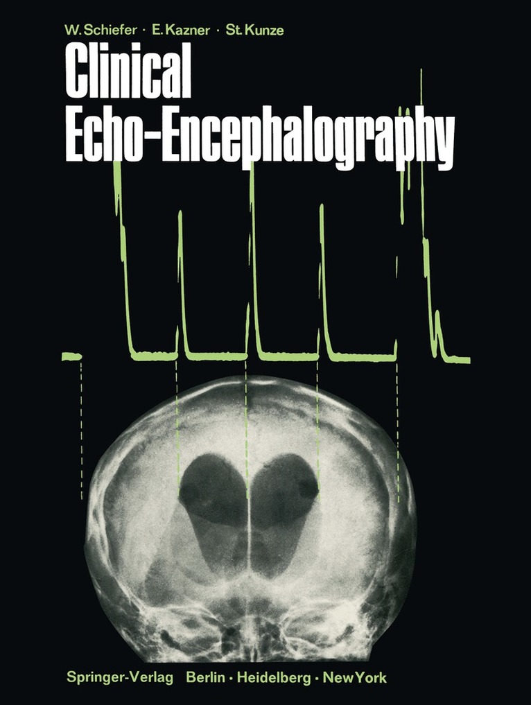
Echo-encephalography, introduced by LEKS ELL in 1955, has gained increasing importance for the early detection of numerous intracranial lesions in the last decade. The main advantage of this diagnostic procedure lies in the fact that it permits a rapid orientation about the spatial relationships within the skull without stressing or endangering the patient. Although this method alone only rarely allows a complete diagnosis, the echo-encephalographic findings always indicate which further diagnostic measures are most suitable for establishing the diagnosis with the greatest accuracy in every case. However, the correct interpretation of an echo-encephalogram is possible only, if the findings which are assumed to be pathological are evaluated in the light of the clinical symptomatology. Since JEPPSSON'S excellent monograph on the origin of the midline echo and its importance for the diagnosis of intracranial expansivities, published in 1961, a great deal of work has gone into the development of echo-encephalography all over the world. For this reason the possibilities of this procedure today go far beyond the mere demonstration of a supratentorial shift. Now we can frequently outline the width of the ventricles exactly and localize tumors or hematomas by means of abnormal reflections. Since a detailed description of the technique, application and present-day diagnostic uses of echo-encephalography has not been available as yet, we undertook to fill this gap in the German literature in 1967 with a monograph summarizing the hitherto existing experience as well as our own extensive case mate rial.
E-bok
PDF, Engelska, 20121 459 kr
Läs direkt efter köp
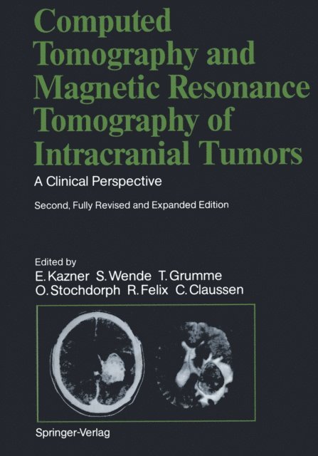
This book represents the second, fully revised edition of the original volume published in 1982. Experience in neuroradiology has confirmed the outstanding value of computed tomography (CT) for the diagnosis of space-occupying lesions within the skull and orbit. It might be assumed, then, that the second edition of this book would simply represent a numerically expanded continua tion of the popular first edition. That is not the case, however. Advances in imaging techniques have promp ted the creation of a new book whose expanded title reflects its more comprehen sive nature. The added illustrations, the revised text, and the expanded circle of editors and contributors document this. Since publication of the first edition, a new modality, magnetic resonance imaging (MRI), has become an established neuroradiologic study. We felt it was essential to include this new modality in our book and explore its capabilities as an adjunct or alternative to CT scanning. Because of the high acquisition costs of MRI and the still small number of MR units currently in operation, we have relied in part on images furnished by other institutions and private practitioners, to whom we are indebted. Many problems relating to MR, both in terms of equipment and image interpretation, have yet to be resolved. There is no denying that we still have much to learn.

Häftad, Engelska, 2011
1 116 kr
Skickas inom 10-15 vardagar
This book represents the second, fully revised edition of the original volume published in 1982. Experience in neuroradiology has confirmed the outstanding value of computed tomography (CT) for the diagnosis of space-occupying lesions within the skull and orbit. It might be assumed, then, that the second edition of this book would simply represent a numerically expanded continua tion of the popular first edition. That is not the case, however. Advances in imaging techniques have promp ted the creation of a new book whose expanded title reflects its more comprehen sive nature. The added illustrations, the revised text, and the expanded circle of editors and contributors document this. Since publication of the first edition, a new modality, magnetic resonance imaging (MRI), has become an established neuroradiologic study. We felt it was essential to include this new modality in our book and explore its capabilities as an adjunct or alternative to CT scanning. Because of the high acquisition costs of MRI and the still small number of MR units currently in operation, we have relied in part on images furnished by other institutions and private practitioners, to whom we are indebted. Many problems relating to MR, both in terms of equipment and image interpretation, have yet to be resolved. There is no denying that we still have much to learn.
E-bok
PDF, Tyska, 2013587 kr
Läs direkt efter köp
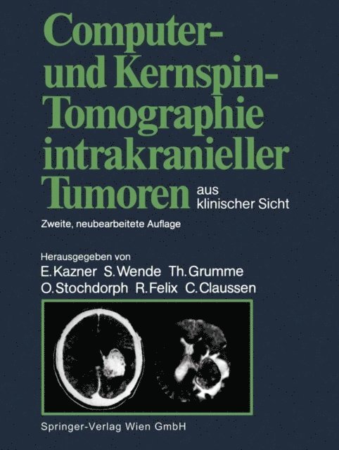
Dieses weltweit bewährte Standardwerk über die zerebrale Computertomographie wurde völlig neu überarbeitet und durch Einbeziehung der Kernspintomographie erweitert. Der Leser erhält eine systematische Darstellung der Diagnose und Differentialdiagnose von nahezu allen Hirngeschwülsten im Computer- und Kernspintomogramm unter Berücksichtigung klinischer Aspekte. Auch Schädelbasis- und Orbita-Prozesse werden mit einbezogen. Grundlage dieses Werkes bildet die umfangreiche Erfahrung der Autoren mit der Computer- und Kernspintomographie bei fast 10.000 Patienten mit verifizierten raumfordernden intrakraniellen Prozessen und Orbita-Erkrankungen. Hervorzuheben ist der umfangreiche Abbildungsteil, in dem nicht nur die häufigsten Geschwulstarten anhand typischer Computer- und Kernspintomogrammen besprochen, sondern auch die seltenen Tumoren und atypischen Lokalisationen demonstriert werden. Für die Einteilung der Tumorgruppen war die neue WHO-Klassifikation maßgebend. In der sehr ausführlichen Differentialdiagnose werden sämtliche nicht tumorbedingten raumfordernden intrakraniellen Prozesse ausführlich erörtert: entzündliche Gehirnerkrankungen, Gehirnerkrankungen im Zusammenhang mit AIDS, akute Entmarkungskrankheiten, Granulome, Zysten, Parasiten, Hirnblutungen, Gefäßmißbildungen und Hirninfarkte.
577 kr
Skickas inom 10-15 vardagar
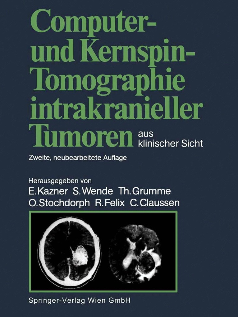
Dieses weltweit bewährte Standardwerk über die zerebrale Computertomographie wurde völlig neu überarbeitet und durch Einbeziehung der Kernspintomographie erweitert. Der Leser erhält eine systematische Darstellung der Diagnose und Differentialdiagnose von nahezu allen Hirngeschwülsten im Computer- und Kernspintomogramm unter Berücksichtigung klinischer Aspekte. Auch Schädelbasis- und Orbita-Prozesse werden mit einbezogen. Grundlage dieses Werkes bildet die umfangreiche Erfahrung der Autoren mit der Computer- und Kernspintomographie bei fast 10.000 Patienten mit verifizierten raumfordernden intrakraniellen Prozessen und Orbita-Erkrankungen. Hervorzuheben ist der umfangreiche Abbildungsteil, in dem nicht nur die häufigsten Geschwulstarten anhand typischer Computer- und Kernspintomogrammen besprochen, sondern auch die seltenen Tumoren und atypischen Lokalisationen demonstriert werden. Für die Einteilung der Tumorgruppen war die neue WHO-Klassifikation maßgebend. In der sehr ausführlichen Differentialdiagnose werden sämtliche nicht tumorbedingten raumfordernden intrakraniellen Prozesse ausführlich erörtert: entzündliche Gehirnerkrankungen, Gehirnerkrankungen im Zusammenhang mit AIDS, akute Entmarkungskrankheiten, Granulome, Zysten, Parasiten, Hirnblutungen, Gefäßmißbildungen und Hirninfarkte.
Häftad, Tyska, 1967
577 kr
Skickas inom 10-15 vardagar
E-bok
PDF, Tyska, 2013550 kr
Läs direkt efter köp