
E. Kazner – författare
Visar alla böcker från författaren E. Kazner. Handla med fri frakt och snabb leverans.
15 produkter
15 produkter
Häftad, Tyska, 1978
577 kr
Skickas inom 10-15 vardagar
Die außergewöhnlich große Bedeutung, welche die axiale Computertomo graphie in der Diagnostik der raumfordernden intracraniellen Prozesse innerhalb weniger Jahre erlangte, hat sich naturgemäß auch auf die Neuro-Traumatologie ausgewirkt. So ist nicht nur das Ausmaß traumati scher Hirnschäden in der Akutphase klarer zu beurteilen, sondern auch die Überwachung der posttraumatischen Verläufe ist um vieles exakter und sicherer geworden. Mit zunehmender Erfahrung haben sich aber auch differentialdiagnostische Probleme ergeben. Es ist deshalb zu begrüßen, daß die Verfasser der vorliegenden Studie die Ergebnisse, die sie an dem großen neuro-traumatalogischen Krankengut zweier neurochirurgischer Kliniken gewonnen haben, im Zusammenhang darstellen. Dabei wird einmal der diagnostische Stellenwert der Methode für die traumatisch bedingten Hirnschädigungen herausgearbeitet, zum anderen auf mögliche Fehlinterpretationen der Befunde hingewiesen. Der Vergleich mit invasiven Untersuchungsmethoden wie der Angiographie und der Luftencephalagraphie läßt die Überlegenheit der nicht invasiven Computertomographie, vor allem in der Verlaufsbeobachtung und der Diagnostik von Folgezuständen, erkennen. Der besondere Wert des Buches liegt darin, daß es von Klinikern geschrieben ist, die sich über die therapeutischen Konsequenzen der Untersuchungsmethode im klaren sind. Bei allem Enthusiasmus für die neue Untersuchungsmethode bleibt die Praevalenz der klinischen Befunde deutlich. Wir möchten dem Buch weite Verbreitung wünschen als Leitfaden für alle Ärzte, die sich mit der Diagnostik, Behandlung und Begutachtung von Patienten mit Schädelhirnverletzungen befassen.
Häftad, Tyska, 1981
754 kr
Skickas inom 5-8 vardagar
E-bok
PDF, Engelska, 20121 174 kr
Läs direkt efter köp
Only a few years following its original development by the English physicist G.N. HOUNSFIELD, cranial computerized tomography has proved to be of revolutionary importance for the diagnosis of brain disorders. This is reflected not l~ast by the almost immediate and worldwide ac ceptance of this diagnostic method. Meanwhile, computerized tomography has in addition led to a considerably improved diagnosis of lesions within the orbital region. With the technically advanced systems of the second generation that will soon be available, the method can also be applied to the study of pathological processes of the facial region of the skull and the neck as well. Finally, although at present still at the stage of clinical investigation, the whole-body scanning system will enable investigation of all parts of the body. Stimulated by the successful 1st and 2nd Internalional Symposium on Computerized Tomography held in Hamilton, Bermuda, in ~''arch 1975 and in San Juan, Puerto Rico, in April 1976, we organized a meeting on cranial computerized tomography which took place in ~unich, June 10 to 12, 1976. It was the aim of this symposium to provide basic informa tion as well as to exchange the experiences with the new method.
E-bok
PDF, Engelska, 20121 408 kr
Läs direkt efter köp
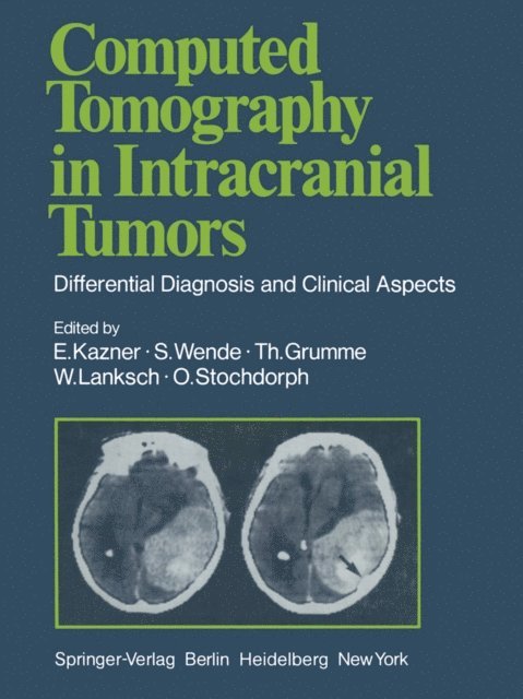
The introduction of computed tomography in the diagnosis of pathological intracranial conditions has had considerable significance in cases of cranio cerebral injury. The decisive diagnostic advantage lies in the possibility of demonstrating both gross pathological change directly as well as secondary changes in normal brain structures. Computed tomography has proved its considerable worth, especially in evaluation of patients with craniocerebral injury and its sequelae. The capabilities of CT were quickly recognized and use of the technique spread rapidly. It is likely that CT will be available within a few years in all hospitals and clinics treating patients with craniocerebral injury. We believe it appropriate to present a detailed report on our experience with CT in 1800 cases of craniocerebral injury treated in the neurosurgical departments in Miinchen-GroBhadern and Berlin-Charlottenburg over a period of five years. Both acute posttraumatic complications and late sequelae are discussed extensively. A large number of illustrations is provided in order to facilitate the reader''s introduction to CT diagnosis. The great interest in our conjoint study originally published in the German language, induced us to translate this book and to update the clinical material. We wish to thank the Stiftung Volkswagenwerk, the Senat of Berlin, the Ludwig-Maximilians-Universitat in Munich and the Freie Universitat of Berlin for the generous financial support which made this study possible.
Häftad, Engelska, 2011
1 115 kr
Skickas inom 10-15 vardagar
The introduction of computed tomography in the diagnosis of pathological intracranial conditions has had considerable significance in cases of cranio cerebral injury. The decisive diagnostic advantage lies in the possibility of demonstrating both gross pathological change directly as well as secondary changes in normal brain structures. Computed tomography has proved its considerable worth, especially in evaluation of patients with craniocerebral injury and its sequelae. The capabilities of CT were quickly recognized and use of the technique spread rapidly. It is likely that CT will be available within a few years in all hospitals and clinics treating patients with craniocerebral injury. We believe it appropriate to present a detailed report on our experience with CT in 1800 cases of craniocerebral injury treated in the neurosurgical departments in Miinchen-GroBhadern and Berlin-Charlottenburg over a period of five years. Both acute posttraumatic complications and late sequelae are discussed extensively. A large number of illustrations is provided in order to facilitate the reader's introduction to CT diagnosis. The great interest in our conjoint study originally published in the German language, induced us to translate this book and to update the clinical material. We wish to thank the Stiftung Volkswagenwerk, the Senat of Berlin, the Ludwig-Maximilians-Universitat in Munich and the Freie Universitat of Berlin for the generous financial support which made this study possible.
E-bok
PDF, Engelska, 20121 459 kr
Läs direkt efter köp
This volume of Advances in Neurosurgery 7 presents the papers held at the Joint Meeting of the American Academy of Neurological Surgery and the "Deutsche Gesellschaft fUr Neurochirurgie" in October 1978 in Munich. This exchange of thoughts on scientific methods in neurosurgery on both sides of the globe, i.e., both in the United States and in Germany, covered a number of different topics in the field of neurosurgery, with special emphasis on the following subjects: Intracranial vascular surgery and specialized neurosurgical techniques used for different operative approaches to the skull, brain, pituitary gland, and peripheral nerves. Contributions to the field of computer tomography, traumatology, functional and experimental neurosurgey, as well as chemotherapy rounded off the broad exchange of thoughts. In particular, the variety of the problems discussed, gives insight into the present state of our special field and shows progress and new points of departure. Special gratitude is expressed to the Springer-\Tedag for its help in editing the Ad vances in Neurosurgery, Volume 7. Miinchen, September 1979 EMARGUTH v Opening Oration F. MARGUTH I should like to welcome all of you wholeheartedly to the Joint Meeting of the American Academy of Neurological Surgery and the Deutsche Gesellschaft fUr N eurochirurgie. I welcome especially our collegues from the United States and the ladies.

Häftad, Engelska, 2011
1 115 kr
Skickas inom 10-15 vardagar
This volume of Advances in Neurosurgery 7 presents the papers held at the Joint Meeting of the American Academy of Neurological Surgery and the "Deutsche Gesellschaft fUr Neurochirurgie" in October 1978 in Munich. This exchange of thoughts on scientific methods in neurosurgery on both sides of the globe, i.e., both in the United States and in Germany, covered a number of different topics in the field of neurosurgery, with special emphasis on the following subjects: Intracranial vascular surgery and specialized neurosurgical techniques used for different operative approaches to the skull, brain, pituitary gland, and peripheral nerves. Contributions to the field of computer tomography, traumatology, functional and experimental neurosurgey, as well as chemotherapy rounded off the broad exchange of thoughts. In particular, the variety of the problems discussed, gives insight into the present state of our special field and shows progress and new points of departure. Special gratitude is expressed to the Springer-\Tedag for its help in editing the Ad vances in Neurosurgery, Volume 7. Miinchen, September 1979 EMARGUTH v Opening Oration F. MARGUTH I should like to welcome all of you wholeheartedly to the Joint Meeting of the American Academy of Neurological Surgery and the Deutsche Gesellschaft fUr N eurochirurgie. I welcome especially our collegues from the United States and the ladies.
E-bok
PDF, Engelska, 20121 459 kr
Läs direkt efter köp
This book represents the second, fully revised edition of the original volume published in 1982. Experience in neuroradiology has confirmed the outstanding value of computed tomography (CT) for the diagnosis of space-occupying lesions within the skull and orbit. It might be assumed, then, that the second edition of this book would simply represent a numerically expanded continua tion of the popular first edition. That is not the case, however. Advances in imaging techniques have promp ted the creation of a new book whose expanded title reflects its more comprehen sive nature. The added illustrations, the revised text, and the expanded circle of editors and contributors document this. Since publication of the first edition, a new modality, magnetic resonance imaging (MRI), has become an established neuroradiologic study. We felt it was essential to include this new modality in our book and explore its capabilities as an adjunct or alternative to CT scanning. Because of the high acquisition costs of MRI and the still small number of MR units currently in operation, we have relied in part on images furnished by other institutions and private practitioners, to whom we are indebted. Many problems relating to MR, both in terms of equipment and image interpretation, have yet to be resolved. There is no denying that we still have much to learn.

Häftad, Engelska, 2011
1 115 kr
Skickas inom 10-15 vardagar
This book represents the second, fully revised edition of the original volume published in 1982. Experience in neuroradiology has confirmed the outstanding value of computed tomography (CT) for the diagnosis of space-occupying lesions within the skull and orbit. It might be assumed, then, that the second edition of this book would simply represent a numerically expanded continua tion of the popular first edition. That is not the case, however. Advances in imaging techniques have promp ted the creation of a new book whose expanded title reflects its more comprehen sive nature. The added illustrations, the revised text, and the expanded circle of editors and contributors document this. Since publication of the first edition, a new modality, magnetic resonance imaging (MRI), has become an established neuroradiologic study. We felt it was essential to include this new modality in our book and explore its capabilities as an adjunct or alternative to CT scanning. Because of the high acquisition costs of MRI and the still small number of MR units currently in operation, we have relied in part on images furnished by other institutions and private practitioners, to whom we are indebted. Many problems relating to MR, both in terms of equipment and image interpretation, have yet to be resolved. There is no denying that we still have much to learn.
E-bok
PDF, Tyska, 2013603 kr
Läs direkt efter köp
E-bok
PDF, Engelska, 20121 459 kr
Läs direkt efter köp
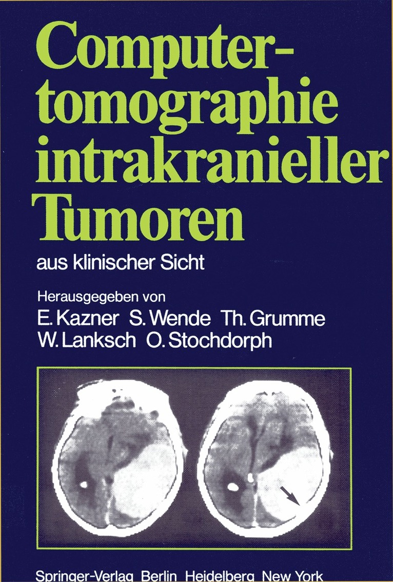
The current book represents a distillation of the experience gained in diagnosis of intracranial tumors with computed X-ray tomography at the University Hos pitals of Berlin, Mainz, and Miinchen. To what purpose? Standard radiological techniques such as pneumoencephalography with lumbar puncture and cerebral arteriography with puncture of the common carotid artery are invasive proce dures which entail a certain amount of risk as well as discomfort for the patient. Furthermore, diagnoses made with these procedures rely primarily on indirect signs of an intracranial space-occupying lesion - such as displacement of the air-filled ventricles or of normal cerebral vessels. Only a few types of tumor are demonstrated directly with these techniques. In contrast, computed tomography demonstrates the pathology directly in almost all cases, and this with a minimum of risk and discomfort. In addition, normal intracranial structures are demonstrated, so that the tumor''s effect on its surroundings can be evaluated. Today, almost a decade after HOUNSFIELD''S revolutionary invention, diagno sis of brain tumors without computed tomography is almost unthinkable, if not in fact irresponsible.

Häftad, Engelska, 2012
1 482 kr
Skickas inom 5-8 vardagar
The current book represents a distillation of the experience gained in diagnosis of intracranial tumors with computed X-ray tomography at the University Hos pitals of Berlin, Mainz, and Miinchen. To what purpose? Standard radiological techniques such as pneumoencephalography with lumbar puncture and cerebral arteriography with puncture of the common carotid artery are invasive proce dures which entail a certain amount of risk as well as discomfort for the patient. Furthermore, diagnoses made with these procedures rely primarily on indirect signs of an intracranial space-occupying lesion - such as displacement of the air-filled ventricles or of normal cerebral vessels. Only a few types of tumor are demonstrated directly with these techniques. In contrast, computed tomography demonstrates the pathology directly in almost all cases, and this with a minimum of risk and discomfort. In addition, normal intracranial structures are demonstrated, so that the tumor's effect on its surroundings can be evaluated. Today, almost a decade after HOUNSFIELD'S revolutionary invention, diagno sis of brain tumors without computed tomography is almost unthinkable, if not in fact irresponsible.

E-bok
PDF, Engelska, 20121 408 kr
Läs direkt efter köp
The investigation of the brain by means of ultrasound has acquired increasing importance in the last years because it permits insight into the spatial relationships within the intact human skull in a short time without endangering the patient. The road from the first ultra sonic investigations on the exposed brain to the detection of intracranial midline shifts on the intact skull, the registration of echo pulsations and recently, to ultrasonotomography has been a long one already. However, this development is by no means at an end. Following the suggestion of numerous colleagues concerned with echo-encephalography in this country and abroad, the Neurosurgical Clinic of the University of Erlangen-Nuremberg organized an "International Symposium on Echo-Encephalography" on April 14th and 15th, 1967. Here there was an open exchange of experience on the results obtained up to the present. The limitations of the method and sources of error as well as the directions of future development of the ultrasonic echo procedure were discussed.

Häftad, Engelska, 2012
1 115 kr
Skickas inom 10-15 vardagar
The investigation of the brain by means of ultrasound has acquired increasing importance in the last years because it permits insight into the spatial relationships within the intact human skull in a short time without endangering the patient. The road from the first ultra sonic investigations on the exposed brain to the detection of intracranial midline shifts on the intact skull, the registration of echo pulsations and recently, to ultrasonotomography has been a long one already. However, this development is by no means at an end. Following the suggestion of numerous colleagues concerned with echo-encephalography in this country and abroad, the Neurosurgical Clinic of the University of Erlangen-Nuremberg organized an "International Symposium on Echo-Encephalography" on April 14th and 15th, 1967. Here there was an open exchange of experience on the results obtained up to the present. The limitations of the method and sources of error as well as the directions of future development of the ultrasonic echo procedure were discussed.
E-bok
PDF, Tyska, 2013587 kr
Läs direkt efter köp
Die außergewöhnlich große Bedeutung, welche die axiale Computertomo graphie in der Diagnostik der raumfordernden intracraniellen Prozesse innerhalb weniger Jahre erlangte, hat sich naturgemäß auch auf die Neuro-Traumatologie ausgewirkt. So ist nicht nur das Ausmaß traumati scher Hirnschäden in der Akutphase klarer zu beurteilen, sondern auch die Überwachung der posttraumatischen Verläufe ist um vieles exakter und sicherer geworden. Mit zunehmender Erfahrung haben sich aber auch differentialdiagnostische Probleme ergeben. Es ist deshalb zu begrüßen, daß die Verfasser der vorliegenden Studie die Ergebnisse, die sie an dem großen neuro-traumatalogischen Krankengut zweier neurochirurgischer Kliniken gewonnen haben, im Zusammenhang darstellen. Dabei wird einmal der diagnostische Stellenwert der Methode für die traumatisch bedingten Hirnschädigungen herausgearbeitet, zum anderen auf mögliche Fehlinterpretationen der Befunde hingewiesen. Der Vergleich mit invasiven Untersuchungsmethoden wie der Angiographie und der Luftencephalagraphie läßt die Überlegenheit der nicht invasiven Computertomographie, vor allem in der Verlaufsbeobachtung und der Diagnostik von Folgezuständen, erkennen. Der besondere Wert des Buches liegt darin, daß es von Klinikern geschrieben ist, die sich über die therapeutischen Konsequenzen der Untersuchungsmethode im klaren sind. Bei allem Enthusiasmus für die neue Untersuchungsmethode bleibt die Praevalenz der klinischen Befunde deutlich. Wir möchten dem Buch weite Verbreitung wünschen als Leitfaden für alle Ärzte, die sich mit der Diagnostik, Behandlung und Begutachtung von Patienten mit Schädelhirnverletzungen befassen.