
H. J. Biersack – författare
Visar alla böcker från författaren . Handla med fri frakt och snabb leverans.
16 produkter
16 produkter
Inbunden, Engelska, 1991
1 231 kr
Skickas inom 10-15 vardagar
Inbunden, Engelska, 1986
1 645 kr
Skickas inom 5-8 vardagar
No detailed description available for "Amphetamines and pH-shift Agents for Brain Imaging".

Inbunden, Engelska, 1995
2 084 kr
Skickas inom 5-8 vardagar
No detailed description available for "Brain SPECT Imaging in Psychiatry".

E-bok
PDF, Engelska, 20191 505 kr
Läs direkt efter köp
No detailed description available for "e;Amphetamines and pH-shift Agents for Brain Imaging"e;.
E-bok
PDF, Engelska, 20151 904 kr
Läs direkt efter köp
No detailed description available for "e;Brain SPECT Imaging in Psychiatry"e;.
Inbunden, Engelska, 2005
1 676 kr
Skickas inom 10-15 vardagar
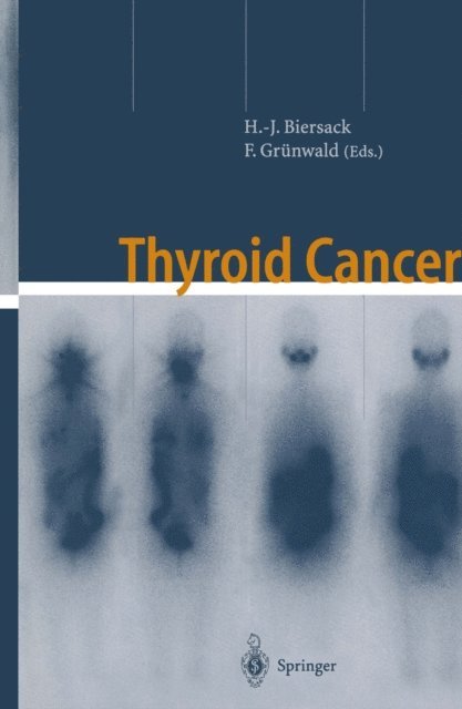
One of the main reasons for publishing this second edition of “T- roid Cancer” is the fact that the first edition has sold out. Furth- more, during the 4 years following the publication of the first edition in 2001, some progress, mainly in the basic sciences (molecular biology), has been made. The most prominent change in the clinical sciences has been the new classification of thyroid cancer, especially with relation to T1–T3 tumors. Now, tumors with a diameter of up to 2 cm are still classified T1. This new UICC classification (6th edition) follows the classification of the American Society of Pathology. These changes require a modification of the old guidelines. According to the Hedinger classification (1988) tumors with a diameter below 1 cm were classified as “papillary microcarcinoma of the thyroid”. Only in those tumors was total or nearly total thyroidectomy deemed unn- essary and I-131 therapy not a prerequisite for treatment. The majority of the chapters has been updated including references to many new publications. Two new chapters, on I-124 PET and - simetry, have been added. We strongly feel that this second edition of “Thyroid Cancer” is again a state-of-the-art overview of the diagnosis and treatment of thyroid cancer. Bonn, Frankfurt am Main H. -J. Biersack, F. Grünwald Preface to the First Edition Thyroid cancer was first described at the end of the eighteenth c- tury.

E-bok
PDF, Engelska, 20051 622 kr
Läs direkt efter köp
One of the main reasons for publishing this second edition of “T- roid Cancer” is the fact that the first edition has sold out. Furth- more, during the 4 years following the publication of the first edition in 2001, some progress, mainly in the basic sciences (molecular biology), has been made. The most prominent change in the clinical sciences has been the new classification of thyroid cancer, especially with relation to T1–T3 tumors. Now, tumors with a diameter of up to 2 cm are still classified T1. This new UICC classification (6th edition) follows the classification of the American Society of Pathology. These changes require a modification of the old guidelines. According to the Hedinger classification (1988) tumors with a diameter below 1 cm were classified as “papillary microcarcinoma of the thyroid”. Only in those tumors was total or nearly total thyroidectomy deemed unn- essary and I-131 therapy not a prerequisite for treatment. The majority of the chapters has been updated including references to many new publications. Two new chapters, on I-124 PET and - simetry, have been added. We strongly feel that this second edition of “Thyroid Cancer” is again a state-of-the-art overview of the diagnosis and treatment of thyroid cancer. Bonn, Frankfurt am Main H. -J. Biersack, F. Grünwald Preface to the First Edition Thyroid cancer was first described at the end of the eighteenth c- tury.
Häftad, Tyska, 2002
527 kr
Skickas inom 10-15 vardagar
Häftad, Engelska, 2014
1 231 kr
Skickas inom 10-15 vardagar
One of the main reasons for publishing this second edition of “T- roid Cancer” is the fact that the first edition has sold out. Furth- more, during the 4 years following the publication of the first edition in 2001, some progress, mainly in the basic sciences (molecular biology), has been made. The most prominent change in the clinical sciences has been the new classification of thyroid cancer, especially with relation to T1–T3 tumors. Now, tumors with a diameter of up to 2 cm are still classified T1. This new UICC classification (6th edition) follows the classification of the American Society of Pathology. These changes require a modification of the old guidelines. According to the Hedinger classification (1988) tumors with a diameter below 1 cm were classified as “papillary microcarcinoma of the thyroid”. Only in those tumors was total or nearly total thyroidectomy deemed unn- essary and I-131 therapy not a prerequisite for treatment. The majority of the chapters has been updated including references to many new publications. Two new chapters, on I-124 PET and - simetry, have been added. We strongly feel that this second edition of “Thyroid Cancer” is again a state-of-the-art overview of the diagnosis and treatment of thyroid cancer. Bonn, Frankfurt am Main H. -J. Biersack, F. Grünwald Preface to the First Edition Thyroid cancer was first described at the end of the eighteenth c- tury.
E-bok
PDF, Tyska, 2013462 kr
Läs direkt efter köp
E-bok
PDF, Engelska, 2012734 kr
Läs direkt efter köp
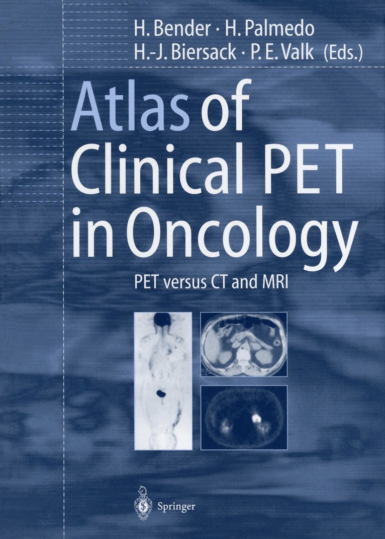
Clinical studies during the past 10 years have shown that PET is more sensitive than CT and MRI for the detection of many tumors. In many cases, however, for example in head and neck tumors, combination with radiological procedures is necessary. It may be speculated that PET should be the first study in a malignant tumor when metastatic spread is suspected. MRI and CT may then be restricted to those body areas which evince sites of increased glucose metabolism. Thus, a combination of metabolic and morphologic procedures will enhance tumor detection and change the therapeutic strategy. In this light, an atlas including PET, CT, MRI, and histology data seems desirable to combine metabolic and morphologic imaging. This book presents an overview of the available data which should be of great interest not only for specialists in radiology and nuclear medicine, but also for oncologists.
E-bok
PDF, Engelska, 20121 459 kr
Läs direkt efter köp
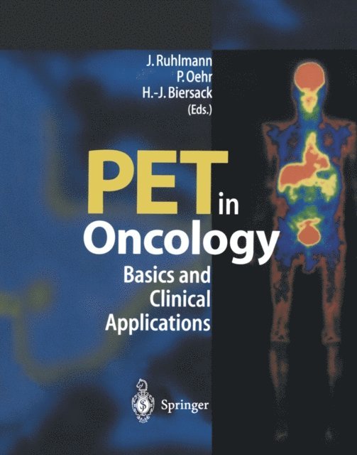
PET in Oncology describes the principles of positron emission tomography and is a useful resource for incorporating the technique in clinical practice. In clear and straightforward fashion, this book offers instructive information and overviews of the physical, biochemical and clinical principles of PET scanning and its routine clinical use. It serves as a reference work for specialists in nuclear medicine and for oncologists, and also provides students and physicians in other medical specialties with a general introduction to the effective integration of this modern technique in routine clinical diagnostics. Above all, this book illustrates the importance of PET in comparison with other imaging techniques.
Häftad, Engelska, 2014
563 kr
Skickas inom 10-15 vardagar
Clinical studies during the past 10 years have shown that PET is more sensitive than CT and MRI for the detection of many tumors. In many cases, however, for example in head and neck tumors, combination with radiological procedures is necessary. It may be speculated that PET should be the first study in a malignant tumor when metastatic spread is suspected. MRI and CT may then be restricted to those body areas which evince sites of increased glucose metabolism. Thus, a combination of metabolic and morphologic procedures will enhance tumor detection and change the therapeutic strategy. In this light, an atlas including PET, CT, MRI, and histology data seems desirable to combine metabolic and morphologic imaging. This book presents an overview of the available data which should be of great interest not only for specialists in radiology and nuclear medicine, but also for oncologists.
Häftad, Engelska, 2011
1 120 kr
Skickas inom 10-15 vardagar
PET in Oncology describes the principles of positron emission tomography and is a useful resource for incorporating the technique in clinical practice. In clear and straightforward fashion, this book offers instructive information and overviews of the physical, biochemical and clinical principles of PET scanning and its routine clinical use. It serves as a reference work for specialists in nuclear medicine and for oncologists, and also provides students and physicians in other medical specialties with a general introduction to the effective integration of this modern technique in routine clinical diagnostics. Above all, this book illustrates the importance of PET in comparison with other imaging techniques.
E-bok
PDF, Tyska, 2013554 kr
Läs direkt efter köp
Es wird erstmals ein kurzgefaßtes Kompendium der Nuklearmedizin für Studenten und Assistenzärzte aller Disziplinen vorgestellt. In allgemeinverständlicher Form werden die wichtigsten nuklearmedizinischen Methoden in das Spektrum anderer Diagnoseverfahren eingeordnet und Indikationen für nuklearmedizinische Untersuchungen herausgearbeitet.
E-bok
PDF, Engelska, 20131 174 kr
Läs direkt efter köp
Thyroid cancer was first described at the end of the eighteenth cen tury. For one and a half centuries surgery remained the only effective therapeutic option for this cancer, until in 1946 radioiodine therapy was performed for the first time. Radioiodine therapy was brought to Germany 4 years later, in 1950. In the intervening 50 years, the use of iodine-131 has proved able to cure the cancer and its metastases. Per cutaneous radiation therapy had been added to the therapeutic ar mamentarium, but even now there is heated debate as to its potential. Suppressive 1-thyroxine supplement is a prerequisite for successful treatment, while cytotoxic drugs are mainly used for palliation. During the past 10 years, various new diagnostic and therapeutic approaches have been introduced. High-dose radioiodine therapy as well as redifferentiation therapy with retinoic acid seem beneficial. Diagnostic procedures such as magnetic resonance imaging (MRI), positron emission tomography (PET), as well as isonitriles (MIBI) 201 and thallium ( Tl), have proved useful for the follow-up of thyroid cancer. Two special issues are also discussed in this book. Iodine supple mentation in areas of iodine deficiency has led to a change in pathol ogy insofar as papillary thyroid cancer (with a better prognosis) has become more frequent than follicular carcinoma. A special chapter is dedicated to thyroid cancer in Chernobyl children.