
Juergen K Mai – författare

3 259 kr
Läs direkt efter köp
2 453 kr
Läs direkt efter köp
2 522 kr
Skickas inom 5-8 vardagar
The fourth edition of Atlas of the Human Brain presents the anatomy of the brain at macroscopic and microscopic levels, featuring different aspects of brain morphology and topography. This greatly enlarged new edition provides the most detailed and accurate delineations of brain structure available. It includes features which assist in the new fields of neuroscience - functional imaging, resting state imaging and tractography. Atlas of the Human Brain is an essential guide to those working with human brain imaging or attempting to relate their observations on experimental animals to humans. Totally new in this edition is the inclusion of Nissl plates with delineation of cortical areas (Brodmann's areas), the first time that these areas have been presented in serial histological sections.
Winner of the 2016 British Medical Association Award for Best Illustrated Text and previous edition winner of the Award of Excellence from the American Association of PublishersThe contents of the Atlas of the brain in MNI stereotaxic space has been extensively expanded from 143 pages, showing 69 levels through the hemisphere, to 314 pages representing 99 levelsIn addition to the fiber-stained (myelin) plates, we now provide fifty new (Nissl) plates covering cytoarchitecture. These are interdigitated within the existing myelin plates of the stereotaxic atlasAll photographic plates now represent the complete hemisphereAll photographs of the cell- and fiber-stained sections have been transformed to fit the MNI-spaceMajor fiber tracts are identified in the fiber-stained sectionsIn the Nissl plates cortical delineations (Brodmann’s areas) are provided for the first timeThe number of diagrams increased to 99. They were now generated from the 3D reconstruction of the hemisphere registered to the MNI- stereotaxic space. They can be used for immediate comparison between our atlas and experimental and clinical imaging resultsParts of cortical areas are displayed at high magnification on the facing page of full page Nissl sections. Images selected highlight those areas which are thought to correspond with those published by von Economo and Koskinas (1925)A novel way of depicting cortical areal pattern is used: The cortical cytoarchitectonic ribbon is unfolded and presented linearly. This linear representation of the cortex enables the comparison of different interpretations of cortecal areas and allows mapping of activation sitesLow magnification diagrams in the horizontal (axial) and sagittal planes are included, calculated from the 3D model of the atlas brain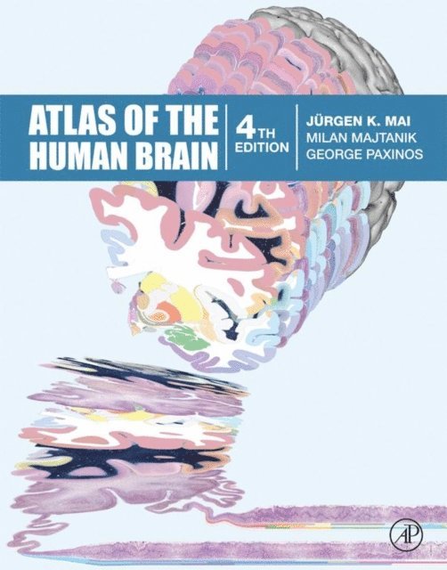
2 759 kr
Läs direkt efter köp
1 020 kr
Skickas inom 10-15 vardagar
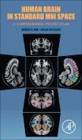
Human Brain in Standard MNI Space: A Comprehensive Pocket Atlas is a thorough pocket atlas designed for easy reference and interpretation of medical and scientific MR-images. It is intended for both early career and advanced medical students, for residents in radiology and neurology, and those involved in neuroscience research, emphasizing anatomy's relationship to radiology.
In addition, the book is ideal for non-specialists interested in issues relating to the brain or the determination of imaging features.
Provides gyral/sulcal designations (in the MNI figures), as well as cortical (Brodmann's areas) delineations (in the diagrams) Contains a three page section with (small) diagrams, providing 3D reconstruction of the MNI brain with definition of the cortex gyri and sulci Includes a section that explains the Brodmann areas, along with a list of abbreviations, structures, and a hierarchical tree of structures
1 448 kr
Läs direkt efter köp

2 422 kr
Kommande

4 177 kr
Kommande