Manfred Tschabitscher – författare
Visar alla böcker från författaren Manfred Tschabitscher. Handla med fri frakt och snabb leverans.
9 produkter
9 produkter
2 714 kr
Skickas inom 7-10 vardagar
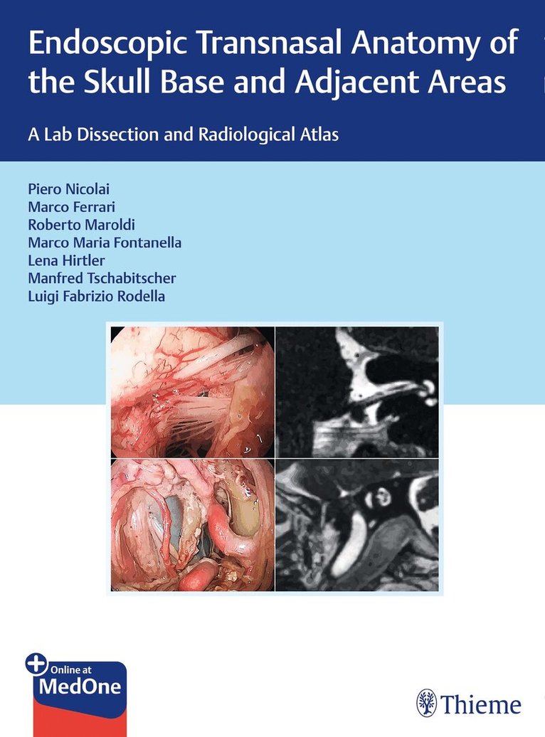
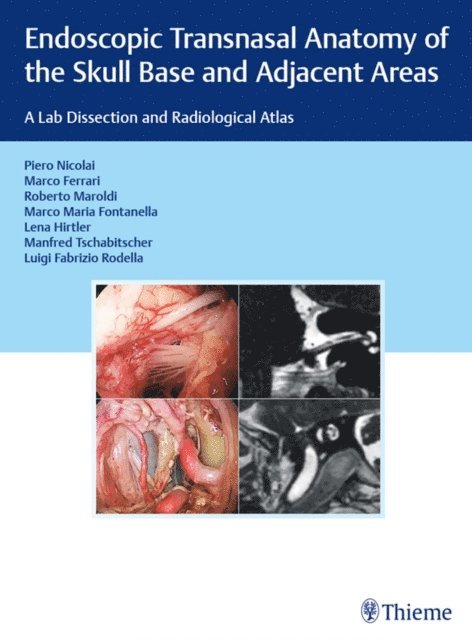
Become familiar with the key anatomic "corridors" in the skull base, the sinonasal tract, and adjacent areas to guide and greatly expand your endoscopic surgical competence.Highlighting the most recent experience from seven top leaders and innovators in the field, this seminal new work presents detailed topographic anatomy of the skull base and adjacent areas in a way not previously seen before.The result is a multidisciplinary atlas merging anatomy, otolaryngology, neurosurgery, and radiology, so as to facilitate creation of a mental "virtual reconstruction" of the complete approach and operative situs. The result is a greatly extended range of surgical possibilities into previously uncharted territory using endoscopic technology.Key Features:Provides the basis for cultivating a firm and confident understanding of the 3D anatomy of this intricately complex regionEmphasizes the ability of the endoscopic surgeon to integrate CT and MRI findings into the surgical planning processA logical and modular organization of the contents intends to make for easy correlation with the surgical literatureBrilliant step-by-step presentation of dissections using cadavers, helping readers to fully understand all the anatomical nuancesNumerous previously unpublished approaches covered here for the first time in a book, step by stepEndoscopic Transnasal Anatomy of the Skull Base and Adjacent Areas is an indispensable resource for fellows and specialists in neurosurgery and ENT surgery wishing to widen their competence in endoscopic skull base surgery.
2 180 kr
Skickas inom 10-15 vardagar
For the beginner or for the accomplished sinus surgeon, mastering the anatomy of the lateral nasal wall is an ongoing challenge. Even though there are excellent standard anatomical references and equally outstanding sinus courses with cadaver dissection, a reference depicting the surgical anatomy is needed. A step-by-step surgical approach on the anterior nasal spine to the anterior wall of the sphenoid is presented. The sinus surgeon is confronted with a wide range of different spaces created by the ethmoid bone. No other bone in the human body has so many anatomic variations. Four critical anatomical structures are emphasized as the foundation for a precise approach to surgery of the maxillary, anterior ethmoid, frontal, and posterior ethmoid sinuses. The goal of this book is to meet the tremendous challenge of offering an anatomical approach which will serve the sinus surgeon of every level of experience and expertise.
1 637 kr
Skickas inom 10-15 vardagar
While microsurgical techniques have already become common knowledge in ENT surgery, a new era has begun with the development of endoscopic surgery. Thereby, however, structures on the opposite side are seen best, not those which have already been examined by the endoscope. One solution to this problem is a biportal approach to the middle ear. Filling an essential gap in anatomical and ENT literature, this atlas shows the various approaches to the middle ear, e.g. , through the tympanic membrane or through the Eustachian tube, which allow safe surgical manipulations.
1 094 kr
Skickas inom 10-15 vardagar
It is only recently that the use of the endoscope as the sole visualizing tool has been introduced in transsphenoidal pituitary surgery with its favorable related implications and minimal operative trauma. Of course, microscopic and endoscopic anatomy are basically the same, but the optical distorsion of endoscopic images is quite substantial compared with microscopic depictions. An endoscope lens produces images with maximal magnification at its center and severe contraction at its periphery. Nearer images are disproportionally enlarged and remote images are falsely miniaturized. This optical illusion may disorientate a surgeon who is not familiar with this peculiar condition at the skull base. This atlas acts as a guide through the endoscopic anatomy and gives detailed descriptions of the preoperative management and the surgical procedures.
Surgical Anatomy of the Internal Carotid Artery
An Atlas for Skull Base Surgeons
Inbunden, Engelska, 2013
1 094 kr
Skickas inom 10-15 vardagar
This atlas provides all the basic and advanced information required by surgeons in order to understand fully the skull base anatomy. It is organized according to anatomo-surgical pathways to the hidden areas of the skull base. These pathways are described in step-by-step fashion with the aid of a wealth of color images and illustrations. The emphasis is on endoscopic anatomy, but in order to provide a holistic perspective, informative three-dimensional reconstructions are presented alongside the endoscopic images and radiologic images are included when appropriate. In effect, windows are opened on the anatomy so that the reader is guided on a journey throughout the skull base region. This anatomically oriented atlas will serve as an ideal learning tool for novice surgeons and will also prove an invaluable reference for the more experienced surgeon
Surgical Anatomy of the Internal Carotid Artery
An Atlas for Skull Base Surgeons
Häftad, Engelska, 2016
1 311 kr
Skickas inom 10-15 vardagar
This atlas provides all the basic and advanced information required by surgeons in order to understand fully the skull base anatomy. It is organized according to anatomo-surgical pathways to the hidden areas of the skull base. These pathways are described in step-by-step fashion with the aid of a wealth of color images and illustrations. The emphasis is on endoscopic anatomy, but in order to provide a holistic perspective, informative three-dimensional reconstructions are presented alongside the endoscopic images and radiologic images are included when appropriate. In effect, windows are opened on the anatomy so that the reader is guided on a journey throughout the skull base region. This anatomically oriented atlas will serve as an ideal learning tool for novice surgeons and will also prove an invaluable reference for the more experienced surgeon
1 637 kr
Skickas inom 10-15 vardagar
if indications are defined by stringent criteria, the technique will gain an impor tant place particularly in pediatric otology. Its clinical indications can naturally not be covered in this volume. As the working space available is limited and the structures in it are very deli cate, the iatrogenic trauma associated with endoscopy must be minimized at all cost. This requires a profound understanding of anatomy. This volume will help the readers acquire or broaden it. April 2000 M. Tschabitscher C.Klug VI Acknowledgements The long tradition of the Institute of Anatomy at the University of Vienna to collaborate with clinical services, which was begun by HYRTL and continued by ZUCKERKANDL, TANDLER, PERNKOPF, v. HAYEK, GISEL, KRAUSE and FERNER, gene rated an atmosphere which still is singularly conducive to research in gross anatomy. We gratefully acknowledge the generous support of our work by the Medical School of the University of Vienna. But we are also indebted to AESCULAP, STORZ and ZEPPELIN companies for providing us with instruments. Our special thanks go to TONI NOBLES from NOBLES-LAI INC., who made the Hawk Vision System with its ultrathin flexible scopes available to us. Without it endoscopic work of this quality would not have been possible. With gratitude we recognize the assistance of Balas Fabinyi, M.D., semor resident at the ENT Service, Krems Hospital, in clinical matters. We are very grateful to Mag. Krista Schmidt for preparing the English-language manuscripts.
1 094 kr
Skickas inom 10-15 vardagar
It is only recently that the use of the endoscope as the sole visualizing tool has been introduced in transsphenoidal pituitary surgery with its favorable related implications and minimal operative trauma. Of course, microscopic and endoscopic anatomy are basically the same, but the optical distorsion of endoscopic images is quite substantial compared to microscopic depictions. An endoscope lens produces images with maximal magnification at its center and severe contraction at its periphery. Nearer images are disproportionally enlarged and remote images are falsely miniaturized. This optical illusion may disorientate a surgeon who is not familiar with this peculiar condition at the skull base. This atlas acts as a guide through the endoscopic anatomy and gives detailed descriptions of the preoperative management and the surgical procedures.
2 180 kr
Skickas inom 10-15 vardagar
For the beginner or for the accomplished sinus surgeon, mastering the anatomy of the lateral nasal wall is an ongoing challenge. Even though there are excellent standard anatomical references and equally outstanding sinus courses with cadaver dissection, a reference depicting the surgical anatomy is needed. A step-by-step surgical approach on the anterior nasal spine to the anterior wall of the sphenoid is presented. The sinus surgeon is confronted with a wide range of different spaces created by the ethmoid bone. No other bone in the human body has so many anatomical variations. Four critical anatomical structures are emphasized as the foundation for a precise approach to surgery of the maxillary, anterior ethmoid, frontal, and posterior ethmoid sinuses. The goal of this book is to meet the tremendous challenge of offering an anatomical approach which will serve the sinus surgeon of every level of experience and expertise.