Enrico de Divitiis – författare
Visar alla böcker från författaren Enrico de Divitiis. Handla med fri frakt och snabb leverans.
5 produkter
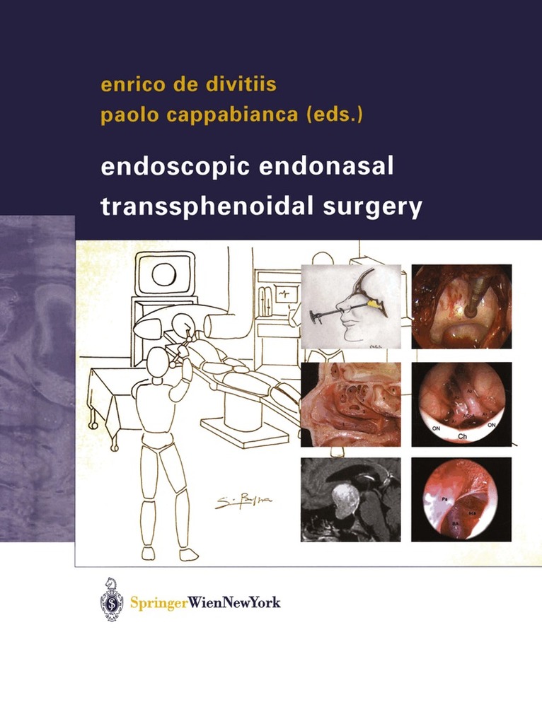
Endoscopic Endonasal Transsphenoidal Surgery
Av Enrico de Divitiis, Paolo Cappabianca
Inbunden, 2003
1686 kr
Lägg i varukorg
Currently, surgical management provides the definitive treatment of choice for most pituitary adenomas, craniopharyngiomas and meningiomas of the sellar region. The elegant minimally invasive transnasal endoscopic approach to the sella turcica and the anterior skull base has added a new dimension …
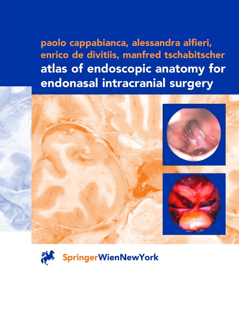
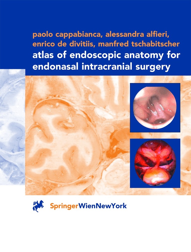
Atlas of Endoscopic Anatomy for Endonasal Intracranial Surgery
Av Paolo Cappabianca, Alessandra Alfieri m. fl.
Inbunden, 2001
1126 kr
Lägg i varukorg
It is only recently that the use of the endoscope as the sole visualizing tool has been introduced in transsphenoidal pituitary surgery with its favorable related implications and minimal operative trauma. Of course, microscopic and endoscopic anatomy are basically the same, but the optical …
Endoscopic Endonasal Transsphenoidal Surgery
Av Paolo Cappabianca, Enrico de Divitiis
E-bok, 2012
2174 kr
Lägg i varukorg
Currently, surgical management provides the definitive treatment of choice for most pituitary adenomas, craniopharyngiomas and meningiomas of the sellar region. The elegant minimally invasive transnasal endoscopic approach to the sella turcica and the anterior skull base has added a new dimension …
Atlas of Endoscopic Anatomy for Endonasal Intracranial Surgery
Av Manfred Tschabitscher, Enrico de Divitiis m. fl.
E-bok, 2012
1470 kr
Lägg i varukorg
It is only recently that the use of the endoscope as the sole visualizing tool has been introduced in transsphenoidal pituitary surgery with its favorable related implications and minimal operative trauma. Of course, microscopic and endoscopic anatomy are basically the same, but the optical …
Atlas of Endoscopic Anatomy for Endonasal Intracranial Surgery
Av Paolo Cappabianca, Alessandra Alfieri m. fl.
Häftad, 2012
1129 kr
Lägg i varukorg
It is only recently that the use of the endoscope as the sole visualizing tool has been introduced in transsphenoidal pituitary surgery with its favorable related implications and minimal operative trauma. Of course, microscopic and endoscopic anatomy are basically the same, but the optical …