
Mukesh G. Harisinghani – författare
816 kr
Skickas inom 10-15 vardagar
1 534 kr
Skickas inom 5-8 vardagar
Widely known as THE survival guide for radiology residents, fellows, and junior faculty, the "purple book" provides comprehensive, up-to-date coverage of diagnostic imaging in an easy-to-read, bulleted format. Focusing on the core information you need for learning and practice, this portable resource combines the full range of diagnostic imaging applications with the latest imaging modalities, making it the perfect clinical companion and review tool.
1 622 kr
Läs direkt efter köp
Detailed anatomic drawings and state-of-the-art radiologic images combine to produce this essential Atlas of Lymph Node Anatomy. Utilizing the most recent advances in medical imaging, this book illustrates the nodal drainage stations in the head and neck, chest, and abdomen and pelvis. Also featured are clinical cases depicting drainage pathways for common malignancies. 2-D and 3-D maps offer color-coordinated representations of the lymph nodes in correlation with the anatomic illustrations. This simple, straightforward approach makes this book a perfect daily resource for a wide spectrum of specialties and physicians at all levels who are looking to gain a better understanding of lymph node anatomy and drainage.
Edited by Mukesh G. Harisinghani, MD, with chapter contributions from staff members of the Department of Radiology at Massachusetts General Hospital.
1 115 kr
Skickas inom 10-15 vardagar
1 459 kr
Läs direkt efter köp
1 226 kr
Skickas inom 10-15 vardagar
816 kr
Skickas inom 10-15 vardagar
1 576 kr
Läs direkt efter köp
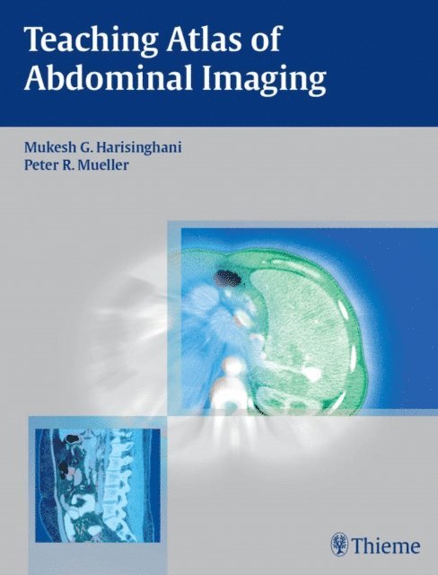
Comprehensive review of diseases of the abdomen and pelvis
Teaching Atlas of Abdominal Imaging is a case-based reference covering the full spectrum of common and uncommon problems of the gastrointestinal and genitourinary tract encountered in everyday practice. The book organizes cases into sections based on the anatomic location of the problem. Each chapter provides succinct descriptions of clinical presentation, radiologic findings, diagnosis, and differential diagnosis for the case. The chapter then discusses the background for each diagnosis, clinical findings, common complications, etiology, imaging findings, treatment, and prognosis.
Key features:
Succinct text and consistent presentation in each chapter enhance the ease of use Practical discussion of all current imaging modalities Nearly 550 high-quality images demonstrate key concepts Bulleted lists of pearls and pitfalls at the end of each chapter highlight important points An appendix with 64-slice protocols for various CT scans, such as dual-phase liver and pancreatic scansIdeal for both self-assessment and rapid review, this book is a valuable resource for radiologists, gastrointestinal and genitourinary radiologists, and fellows and residents in these specialties.
1 669 kr
Skickas inom 10-15 vardagar
1 622 kr
Läs direkt efter köp

949 kr
Skickas inom 10-15 vardagar
1 214 kr
Läs direkt efter köp
This book is a comprehensive atlas on lymph node anatomy and drainage to aid in cancer staging and therapy. Nodal drainage is pertinent to all aspects of cancer staging and therapy and is used by radiation oncologists, surgeons, and medical oncologists to increase accuracy. The first edition of this text was the first comprehensive monograph on this topic, allowing physicians across various specialties to utilize this information and easily share that knowledge with residents, fellows, and junior faculty.
Detailed anatomic drawings and state-of-the-art radiologic images combine to produce this essential Atlas of Lymph Node Anatomy. Utilizing the most recent advances in medical imaging, this book illustrates the nodal drainage stations in the head and neck, chest, abdomen, and pelvis. Also featured are clinical cases depicting drainage pathways for common malignancies. 2-D and 3-D maps offer color-coordinated representations of the lymph nodes in correlation with the anatomic illustrations. This simple, straightforward approach makes this book a perfect daily resource for a wide spectrum of specialties and physicians at all levels who are looking to gain a better understanding of lymph node anatomy and drainage.
This new edition enables physicians to educate themselves on the location of various nodal stations, especially in the context of common primary tumors, so that they are able to detect, localize, and characterize nodes seen with novel new imaging methods and with an increased level of accuracy. Chapters now cover the significant strides made in the imaging realm, such as PET CT, conventional MRI, MRI with novel imaging agents, and multidetector CT, which allows visualization of lymph nodes in various anatomic compartments.
1 447 kr
Skickas inom 10-15 vardagar
1 785 kr
Läs direkt efter köp
This book offers a practical, evidence-based casebook on abdominal imaging. As imaging is increasingly being used for most clinical scenarios, referring physicians rely on radiologists to provide evidence-based interpretation for the incidental and indication related findings seen on the imaging studies. While imagers are trained in identifying the findings, what makes a report more clinically relevant is the accurate assessment and a more focused differential based on knowing the pertinent evidence-based data that supports one diagnosis over the other or favors benign over malignant etiology. Imagers typically don’t have this information readily available to them to provide and make a more informed impression of the findings seen. For example, if a hyperdense renal lesion with attenuation of 80 HU is seen on the noncontrast stone protocol CT, it is important to know what is the likelihood this lesion is worrisome and if any further investigation is needed. While this information is available, it is scattered and one needs to acquire, synthesize, and collate this from the web.
This casebook overcomes this void by providing this information in a simplified, case-based template that any reader can then quickly reference and use in their day-to-day work. In addition, template language for dictation where relevant will be provided. Cases are organized around major abdominal areas, including bowel, genitourinary tract, and pancreas. Each case also includes a table of pearls and pitfalls to be learned and applied to everyday practice.
This is an ideal guide for residents, fellows, and staff from radiology, as a day to day resource or board review book. Given the lack of a comprehensive text, this will be useful to all cadres of radiologists.
1 775 kr
Tillfälligt slut