
Suresh Kumar Mukherji – författare
723 kr
Skickas inom 11-20 vardagar
2 510 kr
Läs direkt efter köp
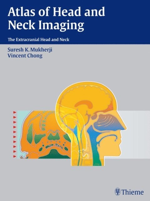
Designed for easy use at the PACS station of viewbox, here is your right-hand tool and pictorial guide for locating, identifying, and accurately diagnosing lesions of the extracranial head and neck. This beautifully produced atlas employs the spaces concept of analysis, which helps radiologists directly visualize complex head and neck anatomy and pathology.
With hundreds of high quality illustrations, this book makes the identification and localization of complex neck masses relatively simple. This book provides CT and MR examples for more than 200 different diseases of the suprahyoid and infrahyoid neck, as well as clear and concise information on the epidemiology, clinical findings, pathology, and treatment guidelines for each disease.
Each space within the head and neck has its own separate section, with examples of the common pathology that arises in this area. A standard format consisting of "Epidemiology, Clinical Presentation, Pathology, Treatment, and Imaging Findings," allows quick and efficient access to well-structured subjects. This uniform organization streamlines research for radiologists at any level of training.
Although well over 200 pathologies are included within this remarkable text, Atlas of Head and Neck Imaging focuses primarily on the suprahyoid and infrahyoid neck, providing exceptionally detailed information on the most challenging aspects of this field.
Radiologists and radiation oncologists will find this visual text ideal as a quick anatomic reference and diagnostic tool. Radiology residents preparing for board exams and neuroradiology fellows and staff studying for the CAQ exam will also benefit from the wealth of information.
1 576 kr
Läs direkt efter köp
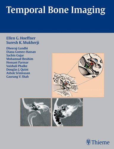
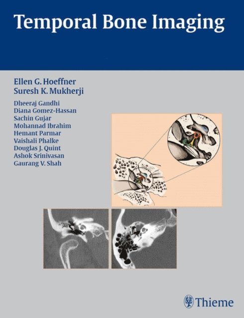
Concise coverage of common temporal bone pathologies in a case-based format
Temporal Bone Imaging is a case-based review of the current techniques for imaging the various temporal bone pathologies frequently encountered in the clinical setting. Detailed discussion of anatomy provides essential background on the complex structure of the temporal bone, as well as the external auditory canal, middle ear and mastoid air cells, facial nerve, and inner ear. Chapters are divided into separate sections based on the anatomic location of the problem, with each chapter addressing a different disease entity.
Highlights:
Each chapter features succinct descriptions of epidemiology, clinical features, pathology, treatment, and imaging findings for CT and MRI Bulleted lists of pearls highlight important imaging considerations More than 200 high-quality images demonstrate anatomy, pathologic concepts, as well as postoperative outcomesThis book will serve as a valuable reference and refresher for radiologists, neuroradiologists, otologists, and head and neck surgeons. Its concise, case-based presentation will help residents and fellows in radiology and otolaryngology-head and neck surgery prepare for board examinations.