• Fri frakt över 249 kr
  • •
  • Snabba leveranser
  • •
  • Billiga böcker
Kundservice

Du är på sajten för privatpersoner.

Företag, bibliotek eller offentlig verksamhet?

Du handlar på classic.bokus.com, där alla dina funktioner finns intakta.
Till classic.bokus.com
Bokus logotyp. Gå till startsidan.
  • Erbjudanden
  • Student
  • Topplistor
  • Barn & ungdom
  • Bokus Play
  • E-böcker
  • Ljudböcker
  • Pocketböcker
  • Spel och pussel

Upp till 25% på utvalda nyheter →

Sidfot

Mina sidor

    Hjälp

    • Kundservice
    • Vanliga frågor och svar
    • Frakt och leverans
    • Retur vid ångerrätt
    • Reklamera vara
    • Betalning
    • Köpvillkor
    • Allmänna villkor
    • Information om webbplatsens tillgänglighet

    Om Bokus

    • Om oss
    • Pressrum
    • För studenter
    • För företag
    • För bibliotek och offentlig verksamhet
    • För leverantörer
    • Hållbarhet

    Populärt

    • Aktuella erbjudanden
    • Presentkort
    • Studentlitteratur
    • Nya böcker
    • Topplistor
    • Signerade böcker
    • Engelska böcker

    Inspiration

    • Boktips
    • BookTok
    • Barnbokskaraktärer
    • Populära författare
    Logotyp för Bokus
    Följ oss på Facebook (extern länk)Följ oss på Instagram (extern länk)Följ oss på YouTube (extern länk)Följ oss på TikTok (extern länk)
    bokus @ CookiesAnpassa cookiesIntegritetspolicyKöpvillkor
    Till Citymail hemsida (extern länk)Till Budbee hemsida (extern länk)Till Postnord hemsida (extern länk)Till Schenker hemsida (extern länk)Till Early Bird hemsida (extern länk)Till Walleys hemsida (extern länk)
    1. Medicin
    2. Andra medicinska specialiteter
    3. Medicinsk bildbehandling

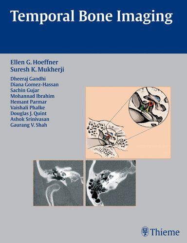
    Temporal Bone Imaging

    AvEllen G. Hoeffner,Suresh Kumar Mukherji

    Inbunden, Engelska, 2008

    751 kr

    Beställningsvara. Skickas inom 11-20 vardagar. Fri frakt över 249 kr.

    Beskrivning

    Concise coverage of common temporal bone pathologies in a case-based formatTemporal Bone Imaging is a case-based review of the current techniques for imaging the various temporal bone pathologies frequently encountered in the clinical setting. Detailed discussion of anatomy provides essential background on the complex structure of the temporal bone, as well as the external auditory canal, middle ear and mastoid air cells, facial nerve, and inner ear. Chapters are divided into separate sections based on the anatomic location of the problem, with each chapter addressing a different disease entity.Highlights: Each chapter features succinct descriptions of epidemiology, clinical features, pathology, treatment, and imaging findings for CT and MRI Bulleted lists of pearls highlight important imaging considerations More than 200 high-quality images demonstrate anatomy, pathologic concepts, as well as postoperative outcomesThis book will serve as a valuable reference and refresher for radiologists, neuroradiologists, otologists, and head and neck surgeons. Its concise, case-based presentation will help residents and fellows in radiology and otolaryngology-head and neck surgery prepare for board examinations.

    Produktinformation

    • Utgivningsdatum:2008-03-26
    • Mått:216 x 279 x 15 mm
    • Vikt:871 g
    • Format:Inbunden
    • Språk:Engelska
    • Antal sidor:244
    • Förlag:Thieme Medical Publishers Inc
    • ISBN:9781588904010

    Utforska kategorier

    • Medicinsk bildbehandling inom Medicin

    Mer om författaren

    Clincal Assistant Professor, Department of Radiology, University of Michigan Health System, Ann Arbor, Michigan, USA

    Recensioner i media

    This is a great book. It is clearly written, concise, complete, and easy to read...An excellent well-priced product.-- RadiologyThe authors do a great job of summarizing the disease process in only 1 or 2 paragraphs...[provides] a good review of each pathologic situation before going into the findings...a great book for both the otolaryngologist and the radiologist, and [the] brevity of each chapter makes it an easy read.--Otology & NeurotologyOrganized by descriptions of diagnoses that make for easy and informative reading...The imaging is crisp and appropriate, and it properly describes the pearls...succinct...a valuable addition to any neuroradiology library (either personal or sectional).--American Journal of Neuroradiology

    Innehållsförteckning

    • Section I Anatomy1 Temporal Bone2 External Auditory Canal3 Middle Ear and Mastoid Air Cells4 Facial Nerve5 Inner EarSection II External Auditory Canal6 External Auditory Canal Atresia and Stenosis7 External Otitis8 Cholesteatoma of the External Auditory Canal9 Exostoses10 External Audiotry Canal Osteoma11 Squamous Cell Carcinoma12 Basal Cell Carcinoma13 MelanomaSection III Middle Ear and Mastoid14 Ossicular Malformations15 Congenital Cholesteatoma16 Aberrant (Intratympanic) Internal Carotid Artery17 Persistent Stapedial Artery18 Dehiscent Jugular Bulb19 Acute Ototis Media and Mastoiditis20 Chronic Otitis Media21 Acquired Cholesteatoma22 Cholesterol Granuloma23 Histiocytosis24 Paraganglioma25 Schwannoma26 Hemangioma27 Meningioma28 Squamous Cell Carcinoma (Middle Ear)29 Adenomatous Lesion30 Adenoid Cystic Carcinoma31 Rhabdomyosarcoma32 MetastasisSection IV Inner Ear and Petrous Bone33 Cochlear Malformations34 Semicircular Canal Dysplasias35 Large Vestibular Aqueduct Syndrome36 Internal Auditory Canal Stenosis/Atresia37 Oval Window Aplasia/Hypoplasia38 Cholesterol Granuloma of the Petrous Apex39 Acute labyrinthitis40 Labyrinthitis Ossifications41 Petrous Apicitis42 Vestibular Schwannoma43 Meningioma44 Congenital Cholesteatoma of the Petrous Apex45 Epidermoid46 Endolymphatic Sac TumorSection V Trauma47 Transverse Temporal Bone Fractures48 Longitudinal Temporal Bone FracturesSection VI Postoperative Ear49 Mastoidectomy50 Ossicular Replacement Prostheses51 Cochlear ImplantationSection VII Miscellaneous52 Otosclerosis53 Fibrous dysplasia54 Paget Disease55 Osteogenesis Imperfecta56 Lymphoma57 Superior Semicircular Canal Dehiscence