Douglas J. Quint – författare
1 219 kr
Läs direkt efter köp
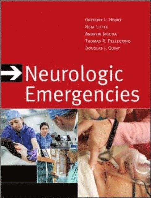
Complete guidance on how to diagnose and treat neurologic emergencies in any acute care setting!
Uniquely organized by presenting complaint, Neurologic Emergencies helps you quickly identify, evaluate, stabilize, treat, and manage the full range of neurologic symptoms--from dizziness to trauma. Striking the perfect balance between thoroughness and readability, this trusted classic delivers the authoritative guidance you need to ensure the best possible outcomes possible for patients presenting with neurologic emergencies. The Third Edition has been updated with a valuable primer on neuroimaging technologies and includes the latest evidence-based treatment strategies and expert advice.
Features:
Organized by presenting symptoms, rather than by disease, for unmatched clinical relevanceWritten by a team of authors highly experienced in managing patients with neurologic complaintsNumerous algorithms and tables offer immediate access to critical informationEmphasizes patient history, physical exam, and rapid assessmentEverything you need to know in the acute care setting:Review of Basic Neuroanatomy, Neuroimaging, Evaluation of a Neurologic Complaint, Altered States of Consciousness and Coma, Acute Focal Neurological Deficit, Acute Weakness, Movement Disorders, Headache; Acute Double Vision, Blindness, and Abnormal Pupils; Neurologic Trauma, Psychogenic Neurologic Problems, Seizures, Syncope, The Dizzy Patient, Neck and Back Pain
723 kr
Skickas inom 11-20 vardagar
1 520 kr
Läs direkt efter köp
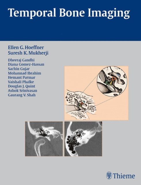
Concise coverage of common temporal bone pathologies in a case-based format
Temporal Bone Imaging is a case-based review of the current techniques for imaging the various temporal bone pathologies frequently encountered in the clinical setting. Detailed discussion of anatomy provides essential background on the complex structure of the temporal bone, as well as the external auditory canal, middle ear and mastoid air cells, facial nerve, and inner ear. Chapters are divided into separate sections based on the anatomic location of the problem, with each chapter addressing a different disease entity.
Highlights:
Each chapter features succinct descriptions of epidemiology, clinical features, pathology, treatment, and imaging findings for CT and MRI Bulleted lists of pearls highlight important imaging considerations More than 200 high-quality images demonstrate anatomy, pathologic concepts, as well as postoperative outcomesThis book will serve as a valuable reference and refresher for radiologists, neuroradiologists, otologists, and head and neck surgeons. Its concise, case-based presentation will help residents and fellows in radiology and otolaryngology-head and neck surgery prepare for board examinations.