
Walter Heindel – författare
2 060 kr
Läs direkt efter köp
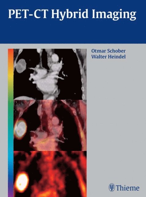
The essentials of PET-CT fusion technology in one practical, comprehensive volume
Written by a multidisciplinary team of experts in radiology and nuclear medicine, this lavishly illustrated handbook presents an evidence-based look at the most up-to-date image-fusion technology in clinical use today. The authors combine multislice spiral CT anatomic images with specific sensitive molecular images of PET in one examination to give readers a full understanding of this evolving technology.
The book places special emphasis on tumor imaging, with additional chapters on the imaging of inflammatory, cardiovascular, and neurodegenerative diseases. Leading clinicians provide systematic discussion of patient preparation, recommendations for imaging protocols for specific indications, and examination techniques such as slice orientation and positioning. To prepare the reader for daily practice, CT and PET-CT scans appear throughout the text side-by-side with explanations of their interpretation.
Highlights:
616 high-quality images - including 175 in full color - complement the text Easy-to-reference textboxes and sidebars present key concepts, pearls, and pitfalls Detailed summaries at the end of each chapter facilitate rapid review Carefully selected suggestions for further reading at the end of each section A comprehensive glossary of frequently used terms and a list of common abbreviationsIdeal for practicing radiologists, radiologic technologists, and radiology residents, PET-CT Hybrid Imaging is an essential reference for anyone who needs to quickly compare and interpret PET-CT images. It is also an excellent preparation tool for Board examinations.
2 612 kr
Läs direkt efter köp
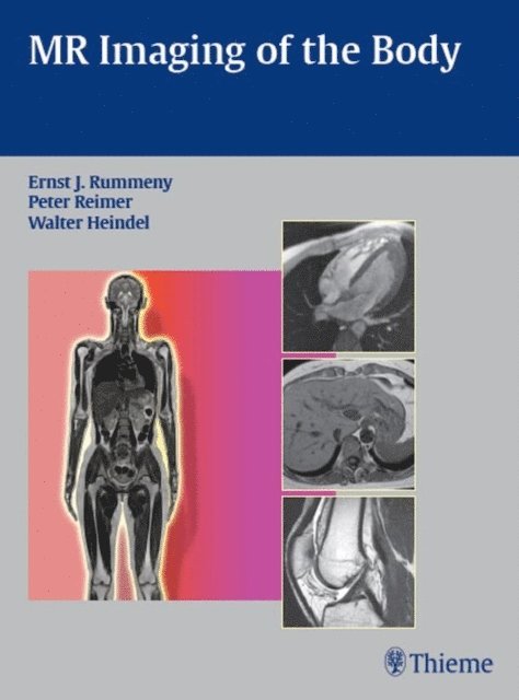
A comprehensive reference for current applications of MR imaging
This book is a comprehensive guide to the basic principles and clinical applications of MR imaging for all regions of the body. The opening chapter provides a thorough overview of the basic principles for MR imaging, contrast agents, risks and side effects associated with MR imaging, and common imaging artifacts. The remaining chapters address common pathological findings in the head and neck, thorax, female breast, abdomen, pelvis, lymph nodes, musculoskeletal system, and vessels. A final chapter discusses the applications of whole-body MRI, whole-body MR angiography, and high-field MRI at 3 T.
Features:
Clear guidelines on how to perform techniques, select the appropriate contrast media, attain the best images, and analyze the findings More than 100 differential diagnosis tables that are ideal for at-a-glance review Detailed comparisons of MR findings and findings using other modalities 1,350 high-quality images and illustrations demonstrating important concepts Bulleted lists of "MRI Specifics" highlighting key pointsThis practical, user-friendly text is a valuable resource for residents and fellows in radiology. It is also an ideal reference for seasoned radiologists seeking to sharpen their MR diagnostic imaging skills.