
Wolfgang Seeger – författare
1 448 kr
Skickas inom 10-15 vardagar

561 kr
Skickas inom 10-15 vardagar

734 kr
Läs direkt efter köp
The main focus of this book is on providing students, neurosurgery trainees, certified neurosurgeons and colleagues in neighbouring disciplines essential information on the evolution of the central nervous system (CNS) of craniata and homo. Therefore the book is divided in three parts: Part I is describing the evolution of CNS of craniata (starting 800 million of years ago). Part II is explaining in detail the exceptional position of the human encephalon. Part III is discussing maturity and immaturity of all parts of CNS of craniatas and the consequences concerning further development of brain structure and psychological functions. In all parts anatomical fundamentals are presented in the form of didactic and self-explanatory illustrations.

561 kr
Skickas inom 10-15 vardagar
1 043 kr
Läs direkt efter köp
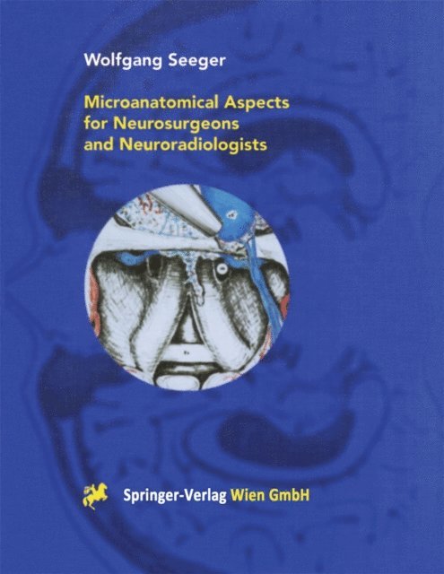
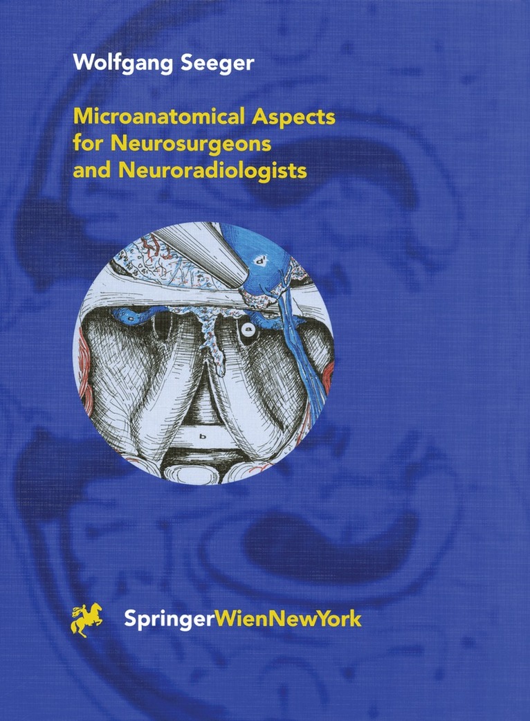
In this atlas anatomical aspects important for combinations of microsurgical and endoscopic approaches are presented and illustrated. Modern imaging techniques are necessary for the three-dimensional orientation but do not show enough details for endoscopic interventions. The small visual fields need a combination of the depiction of fine details and of the three-dimensional presentation of large areas. Furthermore, problems with little known anatomical standard variants of the target areas may arise. Therefore, numerous common anatomical variants are demonstrated with reference to their impact for the surgical technique.
The basis for Professor Seeger’s well known drawings have been anatomical preparations, cadaver dissections and intraoperative pictures. The correct proportions are derived by measuring the distances of anatomical landmarks of cranial preparations and from CT and MR Images. The concise text supports the understanding of the anatomical figures.

2 003 kr
Skickas inom 10-15 vardagar

2 599 kr
Läs direkt efter köp
This detailed atlas illustrates the anatomical structures of the upper basal cisterns, their topography and relationship to other intra- and extradural structures. The author expands his well-established efforts to convey his outstanding neuroanatomical knowledge to the basal cisterns. His famous anatomical drawings are based upon anatomical preparations, cadaver dissections and intraoperative pictures, in order to point out important aspects concerning microsurgical and endoscopic approaches to these parts of the brain.
1 670 kr
Skickas inom 10-15 vardagar
1 579 kr
Läs direkt efter köp

2 003 kr
Skickas inom 10-15 vardagar
2 003 kr
Skickas inom 10-15 vardagar
1 722 kr
Läs direkt efter köp
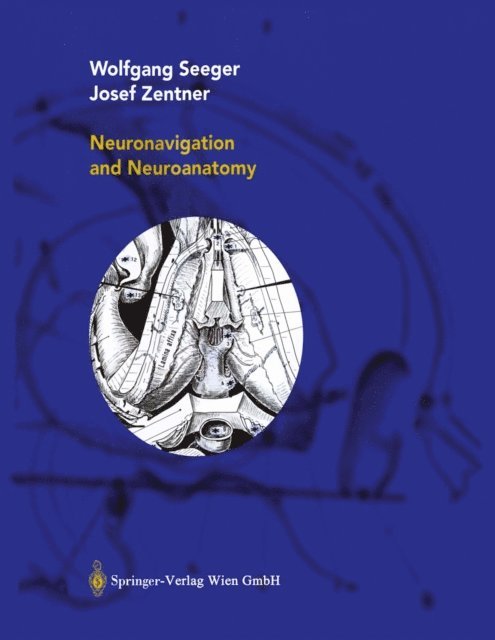
This didactic book clearly and systematically describes the anatomical-surgical fundamentals of cranial neurosurgery, relating them to norm variants, imaging modalities and interdisciplinary aspects. All illustrations, hand drawn in ink by the first author, are simple and self-explanatory.
The book reflects the first author’s lifetime experience as an academic neurosurgeon and teacher, as well as the second author’s theoretical and practical knowledge of neurosurgical subspecialties such as epilepsy surgery.
In addition to its core audience in neurosurgery, it provides all connected disciplines, in particular neuroradiology, neurology, neuropathology, ENT surgery, maxillofacial surgery and eye surgery, with unique anatomical insights into the neurosurgeon’s perspective.
1 459 kr
Läs direkt efter köp

1 116 kr
Skickas inom 10-15 vardagar

554 kr
Läs direkt efter köp

1 227 kr
Skickas inom 10-15 vardagar
1 174 kr
Läs direkt efter köp

1 174 kr
Läs direkt efter köp
577 kr
Skickas inom 10-15 vardagar


734 kr
Läs direkt efter köp
1 132 kr
Läs direkt efter köp
1 785 kr
Läs direkt efter köp
1 132 kr
Läs direkt efter köp

1 174 kr
Läs direkt efter köp
561 kr
Skickas inom 10-15 vardagar
1 337 kr
Skickas inom 10-15 vardagar