• Fri frakt över 249 kr
  • •
  • Snabba leveranser
  • •
  • Billiga böcker
Kundservice

Du är på sajten för privatpersoner.

Företag, bibliotek eller offentlig verksamhet?

Du handlar på classic.bokus.com, där alla dina funktioner finns intakta.
Till classic.bokus.com
Bokus logotyp. Gå till startsidan.
  • Erbjudanden
  • Nyheter
  • Student
  • Topplistor
  • Barn & ungdom
  • Bokus Play
  • E-böcker
  • Pocketböcker
  • Spel & pussel

10% rabatt på allt med kod: NYSTART10 →

Sidfot

Mina sidor

    Hjälp

    • Kundservice
    • Vanliga frågor och svar
    • Frakt och leverans
    • Retur vid ångerrätt
    • Reklamera vara
    • Betalning
    • Köpvillkor
    • Allmänna villkor
    • Information om webbplatsens tillgänglighet

    Om Bokus

    • Om oss
    • Pressrum
    • För studenter
    • För företag
    • För bibliotek och offentlig verksamhet
    • För leverantörer
    • Hållbarhet

    Populärt

    • Aktuella erbjudanden
    • Presentkort
    • Studentlitteratur
    • Nya böcker
    • Topplistor
    • Signerade böcker
    • Engelska böcker

    Inspiration

    • Boktips
    • BookTok
    • Populära bokserier
    • Barnbokskaraktärer
    • Populära författare
    Logotyp för Bokus
    Följ oss på Facebook (extern länk)Följ oss på Instagram (extern länk)Följ oss på YouTube (extern länk)Följ oss på TikTok (extern länk)
    bokus @ CookiesAnpassa cookiesIntegritetspolicyKöpvillkor
    Till Citymail hemsida (extern länk)Till Budbee hemsida (extern länk)Till Postnord hemsida (extern länk)Till Schenker hemsida (extern länk)Till Early Bird hemsida (extern länk)Till Walleys hemsida (extern länk)
    1. Medicin
    2. Andra medicinska specialiteter
    3. Medicinsk bildbehandling

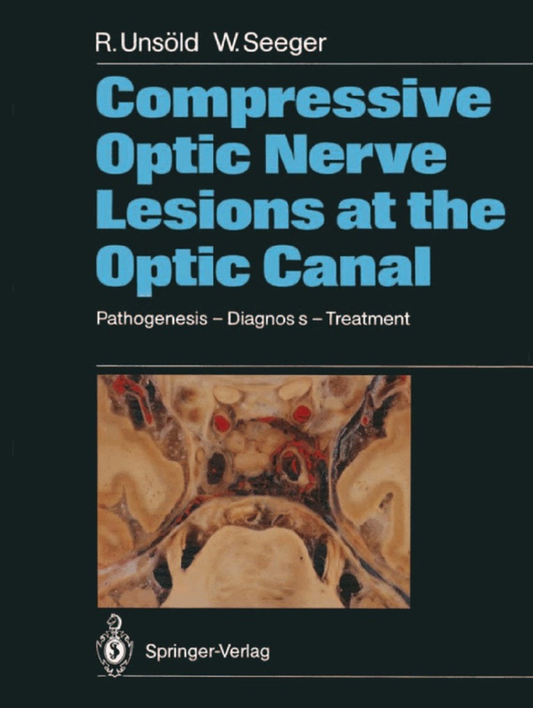
    Compressive Optic Nerve Lesions at the Optic Canal

    Pathogenesis – Diagnosis – Treatment

    AvRenate Unsöld,Wolfgang Seeger

    Häftad, Engelska, 2011

    1 118 kr

    Beställningsvara. Skickas inom 10-15 vardagar. Fri frakt över 249 kr.

    Beskrivning

    The optic canal, in particular its intracranial end, represents a "locus minoris resistentiae" for optic nerve compression in a variety of pathologic conditions. The intracranial optic nerve shares the limited space within this narrow passage with the carotid and ophthalmic artery, all being surrounded by bone and rigid dura. Any pathological condition going along with an increase of soft tissue volume, such as in optic nerve sheath tumors, parasellar neoplasms, dolichoectasia of the carotid and/ or ophthalmic artery, hematomas, etc. , or reduction of the lumen of the bony optic canal by hyperpneumatization of the sphenoid sinus, hyperostosis or developmental abnormalities must act as a space-occupying lesion causing optic nerve compression either by pressing the nerve against the vessel or the neighboring dura or bone.The spectrum of clinical signs and symptoms of optic nerve compression in this area is rather wide and includes acute as well as slowly progressive visual loss and all kinds of visual field defects in the presence of a normal disk, papilledema, pri- mary optic atrophy or cavernous optic atrophy mimicking var- ious clinical disease entities such as retrobulbar optic neuritis, anterior and posterior ischemic optic neuropathy, soft glaucoma and others. Some of the lesions causing optic nerve compression in this area are rather small and need to be visualized or excluded by thin section CT such as pneumosinus dilatans of the sphenoid bone, dolichoectasia of the internal carotid artery, small men- ingiomas around the optic foramen and others.

    Produktinformation

    • Utgivningsdatum:2011-12-06
    • Mått:210 x 280 x undefined mm
    • Vikt:400 g
    • Format:Häftad
    • Språk:Engelska
    • Antal sidor:140
    • Förlag:Springer-Verlag Berlin and Heidelberg GmbH & Co. KG
    • ISBN:9783642733840

    Utforska kategorier

    • Medicinsk bildbehandling inom Medicin
    • Neurokirurgi inom Medicin
    • Oftalmologi inom Medicin

    Innehållsförteckning

    • 1 Introduction.- 2 A Narrow Passage - Anatomic Considerations Topography of the Optic Canal Representing a Predilection of Nerve Compression with Various Pathologic Conditions.- 3 The Clinical Signs and Symptoms of Optic Nerve Compression and Clinical Disease Entities Masking Compressive Lesions.- 3.1 Ophthalmoscopic Findings. Visual Loss, Visual Field Defects, Afferent Pupillary Defect, Color Vision.- 3.2 Visual Evoked Potentials in Optic Nerve Compression.- 4 The Concept of Optic Nerve Compression by Dolichoectatic Arteries Revisited. The Literature and Why It Became Forgotten.- 5 Pneumosinus Dilatans - Rarely Diagnosed and Poorly Understood.- 6 CT Findings of Compressive Lesions at the Optic Canal.- 7 Pterional Approach for Microsurgical Decompression of the Optic Nerve.- 8 Intraoperative Findings in Patients with Intracanalicular Optic Nerve Compression.- 9 Selected Case Reports.- 10 Conclusions.- References.