
Biopsy Pathology Series – serie
Visar alla böcker i serien Biopsy Pathology Series. Handla med fri frakt och snabb leverans.
5 produkter
5 produkter
Häftad, Engelska, 1983
565 kr
Skickas inom 10-15 vardagar
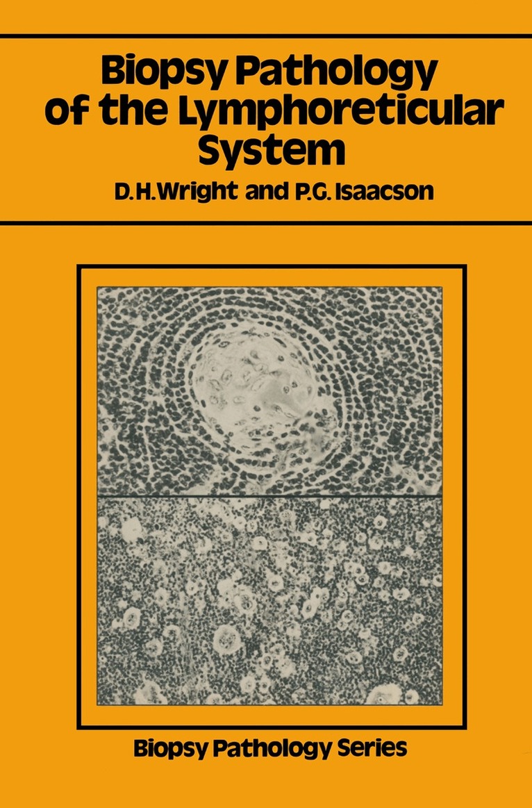
Biopsy pathology of the lymphoreticular system has been written primarily for diagnostic histopathologists although we hope that other workers in the field of lymphoreticular disease will find it of interest and value. With our primary readership in mind we have generously illustrated most sections of the book. Allillustrationsare of haematoxylin and eosin stained sections unless otherwise specified. Conceptual understanding of the histogenesis and interrelationship of non-Hodgkin's Iymphomas has been in a state of turmoil for over a decade. In more recent years immunological and immunocytochemical studies have clarified some problems although in other areas such as the T-celllymphomas histogenetic interrelationships are still far from clear. We are aware, therefore, that in writing this book we have been aiming at a moving target; nevertheless, we feel that the need for such a book, particularly amongst diagnostic histopathologists, outweighs the advan- tage of waiting until all the t's are crossed and all the i's dotted.
Häftad, Engelska, 1986
565 kr
Skickas inom 10-15 vardagar
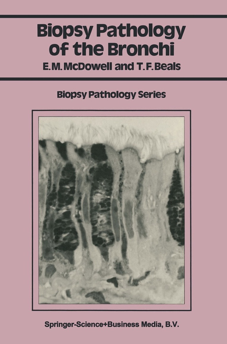
Biopsy Pathology of the Bronchi is intended to provide the practicing pathologist with a convenient source of diagnostic information, combined with pertinent clinical patterns and a background of histogenesis. It is hoped that the book will facilitate the accuracy of diagnosis, minimize pitfalls of interpretation and assist in the rapid evaluation of acute and chronic bronchial lesions, including neoplasms. Biopsy is taken in the widest sense and the book includes discussions on cytology, microbiopsies (fine needle aspiration biopsies, curette samples, and biopsies obtained by microforceps) and macrobiopsies, obtained at the time of surgery. Part I of the book deals with the procurement and preparation of bronchial specimens and includes a chapter on fetal development and structure of the normal adult epithelium. In Part II, the histopathology of numerous bronchial diseases are described in detail. Emphasis is placed on the diagnostic features of human lesions' which are illustrated by numerous high quality light and electron micrographs. Understanding normal bron chial structure, cell kinetics, and modulations of phenotypic expression provide insight into many pathological processes. Therefore, although emphasis is placed on the diagnostic features of human disease, the histopathology of corresponding lesions in experimental animals is dis cussed where this provides information helpful in the interpretation of the human lesions. Elizabeth M. McDowell Baltimore Theodore F. Beals Ann Arbor 1985 viii Acknowledgments We give thanks to friends and colleagues who reviewed one or more chapters for us during the writing of this book.
Häftad, Engelska, 1984
1 123 kr
Skickas inom 10-15 vardagar
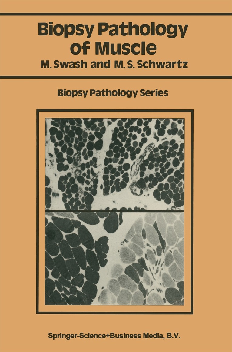
During the last 20 years the development of enzyme histochemical techniques has contributed greatly to knowledge of muscle pathology. However, these and other new methods, such as electron microscopy and immunocytochemistry, have only relatively recently become gener ally available for routine use in histopathology. Muscle biopsy is a long-established technique in clinical practice, having been introduced by Duchenne in 1868 (Arch. Gen. Med. , 11, 5-179). However, the needle method used by Duchenne was not generally adopted, although Shank and Hoagland described a similar technique in 1943 (Science, 98, 592). During this time muscle biopsies required a surgical procedure and this was a considerable disincentive to their use. It was not until Bergstrom (1962; Scand. J. Clin. Lab. Invest. , 14, Suppl. 68) and Edwards (1971; Lancet, ii, 593--6) developed a simple biopsy needle suitable for muscle work in connection with exercise physiology that the advantages of needle muscle biopsies came to be appreciated. Since then, muscle biopsies have become a relatively minor procedure. This has led to the increasing use of muscle biopsy in clinical practice, both for diagnosis and for assessing progress in repeated biopsies during the course of a disorder and its treatment. The full range of enzyme histochemical and ultrastructural histological techniques can be applied to these small biopsies and many of the older histological staining methods can also be used. This book is intended to serve as a practical guide in muscle pathology, particularly for histopathologists, and for those in training.
Häftad, Engelska, 1998
565 kr
Skickas inom 10-15 vardagar
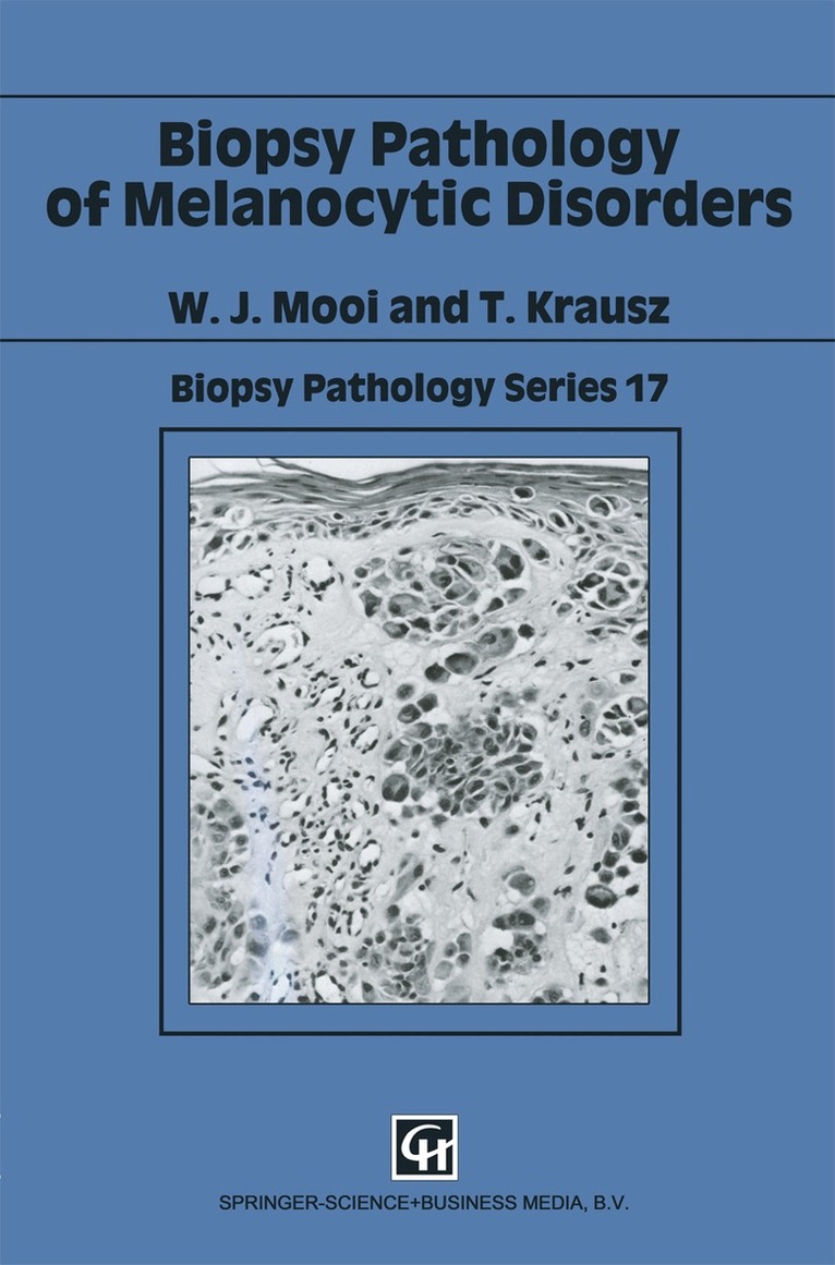
The incidence of cutaneous melanoma has risen considerably over the last few decades, and much attention is now paid to the early diagnosis of this tumour. As a consequence, pathologists are seeing more specimens of pigmented skin lesions. Clinically new entities, such as dysplastic naevi have been identified, and have added to the complexity of the differential diagnosis. Extracutaneous melanocytic lesions are less common but are of considerable clinical importance. This book is a further addition to the "Biopsy Pathology Series", and carries on the tradition of providing a well-illustrated guide for the reporting room pathologist. This has been achieved by highlighting those well-defined histological features which should be used as diagnostic criteria. In addition, tables of diagnostic features are also included, which should facilitate easy reference. This book should be of interest to pathologists and dermatologists.
Häftad, Engelska, 1991
565 kr
Skickas inom 10-15 vardagar
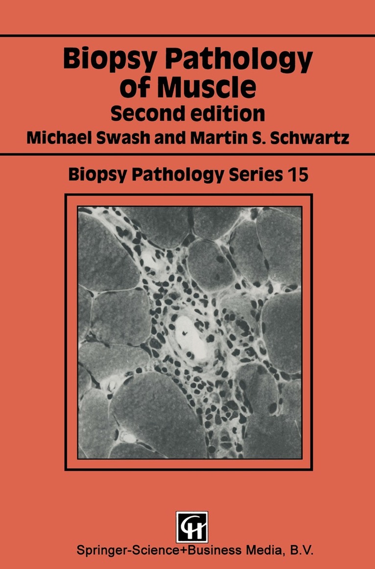
Museie biopsy is a long-established technique in clinical practice having been introduced by Duchenne in 1868 (Arch. Gen. Med. , 11, 5-179). However, the needle method used by Duchenne was not generally adopted, although Shank and Hoagland described a similar technique in 1943 (Science, 98, 592), and open muscle biopsy has for long been preferred in clinical practice, even with the advent of newer needle biopsy methods (Bergstrom, 1962, Scand. J. Clin. Lab. Invest. , 14, Suppl. 68, 1-110). The development of enzyme histochemical techniques has contributed greatly to knowledge of muscle pathology. More recently electron microscopy and immunocytochemistry have also been applied to clinical diagnosis of neuromuscular disease. This book is intended to serve as a practical guide in muscle pathology, particularly for histopathologists, and for those in training. As enzyme histochemistry has become more widely available, formalin-fixed methods have become less frequently used in muscle biopsy work. In this new edition of Muscle Biopsy Pathology we have taken account of the advances in classification and histological technique, and in knowledge of neuromuscular diseases, that have emerged since the first editionwas published in 1984. We hope that this book will continue to be used as a practical guide in the diagnosis and understanding of these disorders. 1. Introduction 1. 1 Generalfeatures of muscle The differentiation of musde into red and white types is a feature of all vertebrates and, indeed, of chordates.