
Martin S. Schwartz – författare
Visar alla böcker från författaren . Handla med fri frakt och snabb leverans.
7 produkter
7 produkter
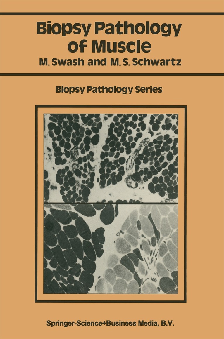
Häftad, Engelska, 1984
1 118 kr
Skickas inom 10-15 vardagar
During the last 20 years the development of enzyme histochemical techniques has contributed greatly to knowledge of muscle pathology. However, these and other new methods, such as electron microscopy and immunocytochemistry, have only relatively recently become gener ally available for routine use in histopathology. Muscle biopsy is a long-established technique in clinical practice, having been introduced by Duchenne in 1868 (Arch. Gen. Med. , 11, 5-179). However, the needle method used by Duchenne was not generally adopted, although Shank and Hoagland described a similar technique in 1943 (Science, 98, 592). During this time muscle biopsies required a surgical procedure and this was a considerable disincentive to their use. It was not until Bergstrom (1962; Scand. J. Clin. Lab. Invest. , 14, Suppl. 68) and Edwards (1971; Lancet, ii, 593--6) developed a simple biopsy needle suitable for muscle work in connection with exercise physiology that the advantages of needle muscle biopsies came to be appreciated. Since then, muscle biopsies have become a relatively minor procedure. This has led to the increasing use of muscle biopsy in clinical practice, both for diagnosis and for assessing progress in repeated biopsies during the course of a disorder and its treatment. The full range of enzyme histochemical and ultrastructural histological techniques can be applied to these small biopsies and many of the older histological staining methods can also be used. This book is intended to serve as a practical guide in muscle pathology, particularly for histopathologists, and for those in training.
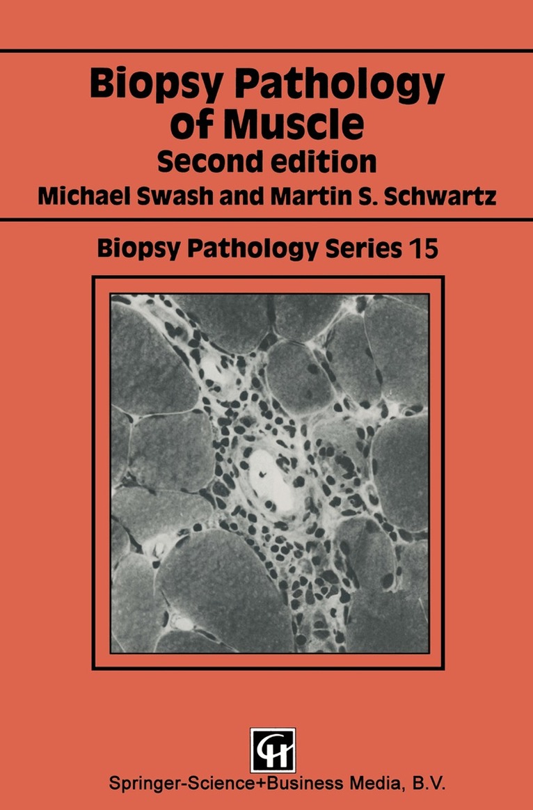
Häftad, Engelska, 1991
563 kr
Skickas inom 10-15 vardagar
Museie biopsy is a long-established technique in clinical practice having been introduced by Duchenne in 1868 (Arch. Gen. Med. , 11, 5-179). However, the needle method used by Duchenne was not generally adopted, although Shank and Hoagland described a similar technique in 1943 (Science, 98, 592), and open muscle biopsy has for long been preferred in clinical practice, even with the advent of newer needle biopsy methods (Bergstrom, 1962, Scand. J. Clin. Lab. Invest. , 14, Suppl. 68, 1-110). The development of enzyme histochemical techniques has contributed greatly to knowledge of muscle pathology. More recently electron microscopy and immunocytochemistry have also been applied to clinical diagnosis of neuromuscular disease. This book is intended to serve as a practical guide in muscle pathology, particularly for histopathologists, and for those in training. As enzyme histochemistry has become more widely available, formalin-fixed methods have become less frequently used in muscle biopsy work. In this new edition of Muscle Biopsy Pathology we have taken account of the advances in classification and histological technique, and in knowledge of neuromuscular diseases, that have emerged since the first editionwas published in 1984. We hope that this book will continue to be used as a practical guide in the diagnosis and understanding of these disorders. 1. Introduction 1. 1 Generalfeatures of muscle The differentiation of musde into red and white types is a feature of all vertebrates and, indeed, of chordates.
E-bok
PDF, Engelska, 20131 174 kr
Läs direkt efter köp
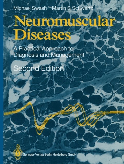
In the seven years since the first edition of this book was published there have been many important developments in knowledge of neuromuscular diseases. These are reflected in this new edition. We have taken the opportunity to add much new clinical and scientific material to the book, particularly in relation to metabolic myopathies and neuropathies, and to include more information on genetic aspects of neuromuscular diseases, quantitative electromyo graphic techniques, plexus and root lesions and cardiomyopathies. The aim of the book remains unchanged, but we have rearranged some of the material so that there are several new chapters. The illustrations have also been extensively revised and there are many new references. We hope that it will continue to provide a convenient source of practical and theoretical information that will not only be useful in managing patients with neuromuscular diseases, but will stimulate research. London, May 1987 Michael Swash Martin S. Schwartz Preface to the First Edition Neuromuscular diseases are common in clinical practice. Patients with these disorders may be referred to neurologists, rheumatologists, orthopaedic surgeons, paediatricians or to general physicians, and their investigation, utilising electromyography (EM G) and muscle biopsy, often requires the help of the clinical neurophysiologist and of the pathologist.

E-bok
PDF, Engelska, 2013734 kr
Läs direkt efter köp
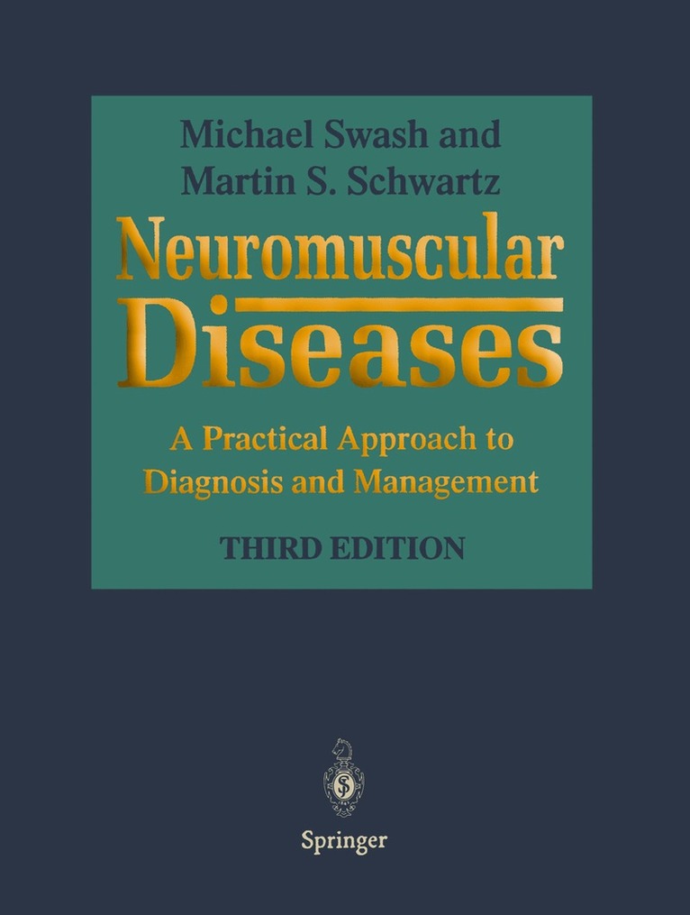
Nine years have elapsed since the second edition of this book was published. In this time the principal advances in neuromuscular diseases have been in the application of molecular genetics to understanding the aetiology and pathogenesis of this group of disorders. As a result many previously unrecognised disorders have been charac terised. Some clinical syndromes, such as the limb girdle dystrophies, have become better defined. In many such instances the new genetic information has led to major advances in knowledge of the biology of cell structures, for example, the membrane structural and channel proteins. The clinical syndromes themselves, and their patho logical and electrophysiological characteristics, however, remain as important as ever, since they constitute the clinical problem itself and, indeed, the database from which all other concepts emerge. Knowledge of the pathogenesis, genetics, and molecular biology of neuromuscular disorders is essential both in developing and applying new therapies and preventive measures, and in formulating genetic and prognostic advice. However, this informa tion does not necessarily always define clinically useful syndromes. Myotonia, for example, is an electrophysiological finding in some syndromes in which it is un detectable by clinical examination, although the phenomenon itself was originally defined as a clinical entity. The limb girdle muscular dystrophy syndromes can be defined by severity, distribution of weakness, age of onset, sex distribution and other characteristics and many of these can be better understood by study of the under lying defect in cell structural proteins.
Häftad, Engelska, 2013
563 kr
Skickas inom 10-15 vardagar
Nine years have elapsed since the second edition of this book was published. In this time the principal advances in neuromuscular diseases have been in the application of molecular genetics to understanding the aetiology and pathogenesis of this group of disorders. As a result many previously unrecognised disorders have been charac terised. Some clinical syndromes, such as the limb girdle dystrophies, have become better defined. In many such instances the new genetic information has led to major advances in knowledge of the biology of cell structures, for example, the membrane structural and channel proteins. The clinical syndromes themselves, and their patho logical and electrophysiological characteristics, however, remain as important as ever, since they constitute the clinical problem itself and, indeed, the database from which all other concepts emerge. Knowledge of the pathogenesis, genetics, and molecular biology of neuromuscular disorders is essential both in developing and applying new therapies and preventive measures, and in formulating genetic and prognostic advice. However, this informa tion does not necessarily always define clinically useful syndromes. Myotonia, for example, is an electrophysiological finding in some syndromes in which it is un detectable by clinical examination, although the phenomenon itself was originally defined as a clinical entity. The limb girdle muscular dystrophy syndromes can be defined by severity, distribution of weakness, age of onset, sex distribution and other characteristics and many of these can be better understood by study of the under lying defect in cell structural proteins.
E-bok
PDF, Engelska, 2013734 kr
Läs direkt efter köp
Museie biopsy is a long-established technique in clinical practice having been introduced by Duchenne in 1868 (Arch. Gen. Med. , 11, 5-179). However, the needle method used by Duchenne was not generally adopted, although Shank and Hoagland described a similar technique in 1943 (Science, 98, 592), and open muscle biopsy has for long been preferred in clinical practice, even with the advent of newer needle biopsy methods (Bergstrom, 1962, Scand. J. Clin. Lab. Invest. , 14, Suppl. 68, 1-110). The development of enzyme histochemical techniques has contributed greatly to knowledge of muscle pathology. More recently electron microscopy and immunocytochemistry have also been applied to clinical diagnosis of neuromuscular disease. This book is intended to serve as a practical guide in muscle pathology, particularly for histopathologists, and for those in training. As enzyme histochemistry has become more widely available, formalin-fixed methods have become less frequently used in muscle biopsy work. In this new edition of Muscle Biopsy Pathology we have taken account of the advances in classification and histological technique, and in knowledge of neuromuscular diseases, that have emerged since the first editionwas published in 1984. We hope that this book will continue to be used as a practical guide in the diagnosis and understanding of these disorders. 1. Introduction 1. 1 Generalfeatures of muscle The differentiation of musde into red and white types is a feature of all vertebrates and, indeed, of chordates.
E-bok
PDF, Engelska, 20131 719 kr
Läs direkt efter köp
During the last 20 years the development of enzyme histochemical techniques has contributed greatly to knowledge of muscle pathology. However, these and other new methods, such as electron microscopy and immunocytochemistry, have only relatively recently become gener ally available for routine use in histopathology. Muscle biopsy is a long-established technique in clinical practice, having been introduced by Duchenne in 1868 (Arch. Gen. Med. , 11, 5-179). However, the needle method used by Duchenne was not generally adopted, although Shank and Hoagland described a similar technique in 1943 (Science, 98, 592). During this time muscle biopsies required a surgical procedure and this was a considerable disincentive to their use. It was not until Bergstrom (1962; Scand. J. Clin. Lab. Invest. , 14, Suppl. 68) and Edwards (1971; Lancet, ii, 593--6) developed a simple biopsy needle suitable for muscle work in connection with exercise physiology that the advantages of needle muscle biopsies came to be appreciated. Since then, muscle biopsies have become a relatively minor procedure. This has led to the increasing use of muscle biopsy in clinical practice, both for diagnosis and for assessing progress in repeated biopsies during the course of a disorder and its treatment. The full range of enzyme histochemical and ultrastructural histological techniques can be applied to these small biopsies and many of the older histological staining methods can also be used. This book is intended to serve as a practical guide in muscle pathology, particularly for histopathologists, and for those in training.