
Medicinsk bildbehandling
Perrino and Reeves' Practical Approach to Transesophageal Echocardiography
Print + ebook with Multimedia
1 459 kr
Skickas inom 7-10 vardagar

1 428 kr
Skickas inom 10-15 vardagar
1 925 kr
Skickas inom 5-8 vardagar
1 157 kr
Skickas
2 019 kr
Skickas inom 5-8 vardagar
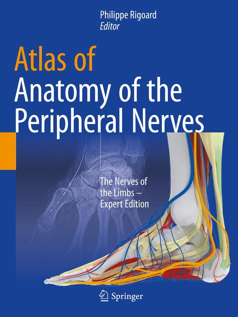
Atlas of Anatomy of the peripheral nerves
The Nerves of the Limbs – Expert Edition
1 963 kr
Skickas inom 10-15 vardagar
1 419 kr
Skickas inom 5-8 vardagar
617 kr
Skickas
Akut radiologi beskriver radiologins roll och möjligheter i vårdkedjan för patienter som söker för akut medicinisk sjukdom eller skada. Texten är anpassad till nordiska förhållanden och täcker de vanligaste akuta tillstånd som kräver avbildning eller intervention.
Boken är indelad i fem delar. Den första handlar om traumatiska tillstånd, den andra om icke-traumatiska tillstånd. De följande behandlar pediatriska akuta tillstånd samt radiologiska undersökningar av intensivvårdspatienter. Slutligen beskrivs radiologiska interventioner och indikationer.
Om författarnaUnder redaktion av;Seppo K. Koskinen, professor och överläkare i bild- och funktionsmedicin vid Karolinska Institutet och Karolinska universitetssjukhuset. Han har en klinisk bakgrund inom sarkom-, trauma- och muskuloskeletal radiologi samt har en omfattande verksamhet inom forskning relaterad till extremitetsskador och multitrauma.
Roberto Blanco Sequeiros, verksamhetschef och professor i Egentliga Finlands Avbildningscentral vid Åbo universitetscentralsjukhus. Han har en klinisk bakgrund inom interventionell och muskuloskeletal radiologi samt har en omfattande verksamhet inom forskning relaterad till cancer och magnetisk resonanstomografi.
Som författare har tolv specialister deltagit, samtliga med stor erfarenhet av klinisk verksamhet inom sina respektive områden.
Sagt om boken"De två redaktörerna är båda professorer i radiologi (röntgen) vid respektive Karolinska universitetssjukhuset och Åbo universitetssjukhus. Tillsammans med andra specialister har de skrivit en alldeles utmärkt bok om akuta radiologiska undersökningar. [...] I texten finns mängder med återgivna röntgenbilder på hur ett flertal akuta tillstånd visar sig på en undersökning. [...]". Publiceras i BTJ-häftet nr 24, 2017. Lektör Lars Svensson
985 kr
Skickas inom 10-15 vardagar
594 kr
Skickas inom 5-8 vardagar
483 kr
Skickas inom 5-8 vardagar
2 256 kr
Skickas inom 7-10 vardagar
1 135 kr
Skickas inom 7-10 vardagar
Workbook and Lab Manual for Sonography
Introduction to Normal Structure and Function
887 kr
Skickas inom 5-8 vardagar
Review important sonography learnings with Curry and Prince's Workbook for Sonography: Introduction to Normal Structure and Function, 5th Edition. This well-constructed review tool supports and completes the main text by providing an excellent introduction to sonography while preparing users to accurately identify sonographic pathology and abnormalities. Each workbook chapter opens with review questions on material from the corresponding chapter in the main text. Review questions are followed by drawings from the text - with parallel sonograms where appropriate - that include leader lines to label structures, but not the labels themselves. Workbook users will fill in the labels to identify structures in the drawings and sonograms, reinforcing visual and auditory learning from the text. Answers can be looked up in both the workbook appendix and by comparing the workbook figures to the labeled figures in the main text.
912 kr
Skickas inom 5-8 vardagar
742 kr
Skickas inom 7-10 vardagar
587 kr
Skickas
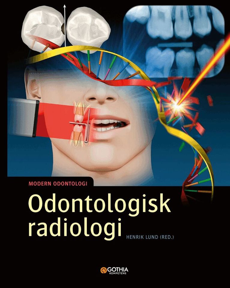
Odontologisk radiologi
Odontologisk radiologi utgör en grundpelare för diagnostik och behandling inom tandvården. Samtidigt innefattar röntgenundersökningen både nytta och risker. Detta ställer höga krav på kunskap om såväl grundläggande röntgenfysik och strålbiologi som inställningsteknik för ett optimalt slutresultat till gagn för patienten.
I boken Odontologisk radiologi beskrivs röntgenstrålningens uppkomst och växelverkan. Författarna går igenom röntgenundersökningens praktiska utförande med målet att åstadkomma en avbildning som ger all nödvändig information med minsta möjliga stråldos. Dessutom beskrivs tolkning av röntgenbilden avseende normalanatomiska strukturer och deras avbildning samt grundläggande röntgendiagnostik med särskilt fokus på folksjukdomarna karies och parodontit.
Boken Odontologisk radiologi vänder sig till studerande på tandsköterske- och tandhygienistutbildningarna men kan också tillämpas inom grundnivå på tandläkarutbildningen samt utgöra en kunskapsaktualisering för hela tandvårdsteamet.
550 kr
Skickas inom 10-15 vardagar
296 kr
Skickas inom 10-15 vardagar
- Nyhet
500 kr
Skickas inom 7-10 vardagar
134 kr
Skickas

Elements of Marie Curie
How the Glow of Radium Lit a Path for Women in Science
261 kr
Skickas
203 kr
Skickas
693 kr
Skickas inom 3-6 vardagar

732 kr
Skickas inom 5-8 vardagar
294 kr
Skickas inom 5-8 vardagar
984 kr
Skickas inom 3-6 vardagar
Publisher's Note: Products purchased from Third Party sellers are not guaranteed by the publisher for quality, authenticity, or access to any online entitlements included with the product.
The Student-to-Student, Step-by-Step Guide to Radiology Clerkship Success
This survival guide for the radiology wards teaches you how to read and present normal and abnormal x-rays, CTs, and MRIs for the most commonly seen diseases and disorders. You'll find 150 radiologic images divided by modality, clear guidelines on when to order a specific modality and how to read the results, and illustrations of classic results of important conditions.
Detailed advice from radiology clerkship veterans help you:
Prepare for and take the test Perform a basic study Determine if a film is adequate Present each study on the ward Spot the strengths and weaknesses of studies Recognize basic anatomical structures Identify abnormal conditionsINSIDER’S GUIDE TO HONING RADIOLOGY SKILLS FOR EVERY CLERKSHIP
The only student-to-student, step-by-step guide to interpreting and presenting radiology studiesDetailed how-to-succeed and what-to-study guidance from clerkship vets75 radiology images of the most commonly seen disorders illustrate each modality and detail Illustrates normal as well as abnormal resultsClassic results of important conditions often seen on the wards and tested on the USMLE Step 2CKClear guidelines on when to order a specific modality and how to read the results“Classifieds” feature high-yield websites, top extracurricular opportunities, and scholarshipsTear-out cards with essential radiology concepts in pocket-ready formatA STUDENT-TO-STUDENT GUIDE
Discover med students’ “secret weapon” Impress on the wards and succeed in the clerkshipsSave time with high-yield topics, important radiology images, and pocket-ready remindersThe first book to describe how to read radiographic films for the most commonly seen disordersApply the First Aid formula for clerkship success!
4 646 kr
Skickas inom 3-6 vardagar
Publisher's Note: Products purchased from Third Party sellers are not guaranteed by the publisher for quality, authenticity, or access to any online entitlements included with the product.
Sharpen your diagnostic skills for brain and spine disorders with this unique patterns-based approach to learning
Brain and Spine Imaging Patterns presents a systematic approach to understanding one of the most challenging areas of radiologic interpretation. Uniquely organized by various patterns seen on CT, MRI, and plain radiography imaging rather than pathology, the book carefully guides you toward a group of differential diagnoses. You will find an unmatched collection of more than 140 patterns covering: skull defects and lesions; meningeal and sulcal diseases; extracerebral masses; intracerebral masses; mass lesions in the region of the ventricular system; sellar and parasellar masses; vascular legions; lesions in the cortical gray matter, white matter, and deep gray matter; and spinal diseases and lesions.
The easy-to-navigate organization of this book is specifically designed for use at the workstation. The concise text, numerous images, and helpful icons facilitate access to essential information and simplify the learning process.
Features
More than 140 patterns and more than 2500 digital-quality imagesA strong focus on patterns recognized on MRI, including contrast-enhanced MRIIcons, a grading system depicting the relative frequency of findings from common to rare, and the consistent organization of chapters help to clarify information for at-the-bench consultationMany patterns include a set of “Related Patterns” images, which serve as cross-references to similar patterns for a given disorder Special emphasis on the latest diagnostic modalities includes state-of-the-art depiction of image findings
3 471 kr
Skickas inom 3-6 vardagar
Publisher's Note: Products purchased from Third Party sellers are not guaranteed by the publisher for quality, authenticity, or access to any online entitlements included with the product.
295 cases and more than 1700 illustrations teach you how to accurately interpret genitourinary tract images
4 STAR DOODY'S REVIEW!"The high-quality images and pithy discussions make this book very useful to radiologists, both in training and in practice....The book's best features are the excellent image quality, the inclusion of images of differential diagnostic considerations, and concise discussions of the cases. This is an excellent resource for radiologists in training and in practice. The case-based format is excellent for board preparation, and its concise prose provides all the necessary information while leaving out the excess."--Doody's Review Service
Genitourinary Imaging Cases presents an efficient and systematic approach to examining images of the genitourinary system. You will find an unmatched collection of 295 cases ranging from normal anatomy to the full spectrum of disease –- including renal cystic masses, renal infection, renal vascular disease, and female pelvic abnormalities. Included with these cases are 1700+ high-quality images that are representative of what you would see on various imaging modalities.
The book’s easy-to-navigate organization is specifically designed for use at the workstation. The concise text, numerous images, and helpful icons speed access to essential information and simplify the learning process.
Features:
Each case includes findings, differential diagnosis, comment/discussion, and clinical pearls Icons, a grading system depicting the full spectrum of diseases, common to rare, and imaging findings, typical to unusual, along with the consistent chapter organization make this perfect for rapid at-the-bench consultation Strong focus on pathology Special emphasis on the latest diagnostic modalities that include both CT and MR images
2 059 kr
Tillfälligt slut
Publisher's Note: Products purchased from Third Party sellers are not guaranteed by the publisher for quality, authenticity, or access to any online entitlements included with the product.
All the clinical applications of whole-body MRI--together in one essential reference!
"This book, to my knowledge, is the first of its kind and offers a hands-on approach to the evolving field of whole-body MRI, which gives the reader tools to effectively utilize and optimize applicatino of whole-body MRI technology in the patient care setting."--F. Scott Pereles, MD, Salinas Valley Radiologists
Superbly illustrated, and written by leading experts who helped establish MRI as a diagnostic tool, this clinically relevant resource puts whole-body MRI into perspective like no other text. The first section covers the technical requirements of whole-body MRI, along with its principles and associated data processing. Subsequent sections offer a practical look at all aspects of whole-body MRI from a clinical perspective--including its use in patients with peripheral arterial occlusive disease and in metastasis screening.
Features:
The only hands-on approach to the evolving field of whole-body MRI-filled with tools to optimize whole-body MRI technology in the patient care settingComplete coverage of all the latest whole-body MRI technical requirements, principles, and associated data processing Richly illustrated presentation of the clinical areas of cardiovascular investigation and musculoskeletal diseasesOverview of oncological whole-body imaging that addresses the comparison of whole-body MRI with bone scintigraphy in the detection of bone metastases, and with FDG-PET as it relates to whole-body stagingDetailed review of both the clinical and experimental questions concerning whole-body MRI