Stefano Fanti – författare
Visar alla böcker från författaren Stefano Fanti. Handla med fri frakt och snabb leverans.
30 produkter
PET Imaging in the Management of Neuroendocrine Tumors, An Issue of PET Clinics
Av Stefano Fanti, Cristina Nanni m. fl.
Inbunden, 2014
840 kr
Lägg i varukorg
Nuclear Medicine has greatly contributed to the diagnosis and treament of neuroendocrine neoplasms. This issue of PET Clinics will focus not only on the diagnosis and treatment of neuroendocrine tumors, but also theranostics. Topics include SPECT and other PET tracers, F-DOPA, Ga-DOTA-peptides, …
Novel PET Imaging Techniques in the Management of Hematologic Malignancies, An Issue of PET Clinics
Av Cristina Nanni, Paolo Castellucci m. fl.
Inbunden, 2024
827 kr
Lägg i varukorg
In this issue of PET Clinics , guest editors Drs. Cristina Nanni, Paolo Castellucci, Stefano Fanti, and Neeta Pandit-Taskar bring their considerable expertise to the topic of Novel PET Imaging Techniques in the Management of Hematological Malignancies . Top experts in the field discuss the latest …
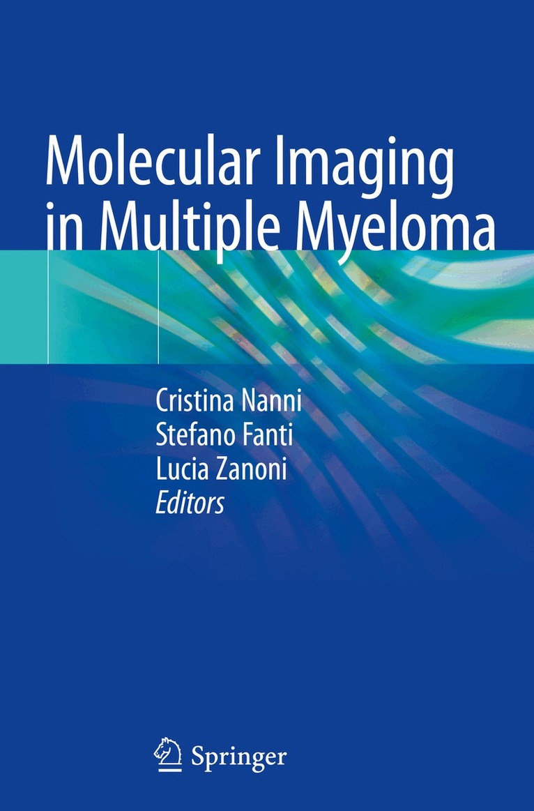
Molecular Imaging in Multiple Myeloma
Av Cristina Nanni, Stefano Fanti m. fl.
Inbunden, 2019
1128 kr
Lägg i varukorg
This book provides a comprehensive overview of the importance of molecular imaging in multiple myeloma, with detailed explanation of its clinical impact. Other important features are the definition of criteria that will aid PET/CT interpretation; identification and explanation of the most frequent …
Molecular Imaging in Multiple Myeloma
Av Lucia Zanoni, Stefano Fanti m. fl.
E-bok, 2019
1251 kr
Lägg i varukorg
This book provides a comprehensive overview of the importance of molecular imaging in multiple myeloma, with detailed explanation of its clinical impact. Other important features are the definition of criteria that will aid PET/CT interpretation; identification and explanation of the most frequent …
Molecular Imaging in Multiple Myeloma
Av Cristina Nanni, Stefano Fanti m. fl.
Häftad, 2020
960 kr
Lägg i varukorg
This book provides a comprehensive overview of the importance of molecular imaging in multiple myeloma, with detailed explanation of its clinical impact. Other important features are the definition of criteria that will aid PET/CT interpretation; identification and explanation of the most frequent …
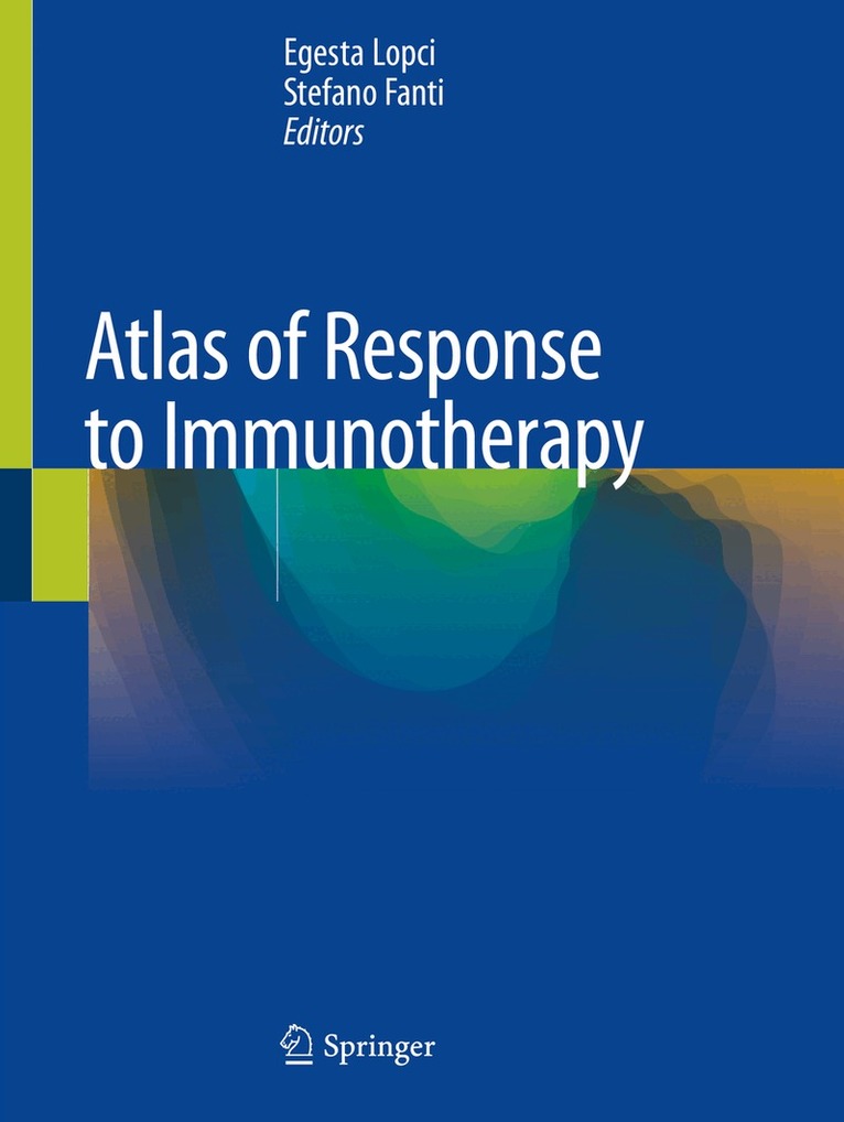
Atlas of Response to Immunotherapy
Av Egesta Lopci, Stefano Fanti
Inbunden, 2019
1688 kr
Lägg i varukorg
This atlas is a concise but comprehensive guide to the diverse patterns of response to immunotherapy as observed on Positron Emission Tomography/Computed Tomography (PET/CT) and other conventional imaging modalities, including CT and Magnetic Resonance Imaging (MRI). The purpose for this …
Atlas of Response to Immunotherapy
Av Stefano Fanti, Egesta Lopci
E-bok, 2019
1670 kr
Lägg i varukorg
This atlas is a concise but comprehensive guide to the diverse patterns of response to immunotherapy as observed on Positron Emission Tomography/Computed Tomography (PET/CT) and other conventional imaging modalities, including CT and Magnetic Resonance Imaging (MRI). The purpose for this …
Atlas of Response to Immunotherapy
Av Egesta Lopci, Stefano Fanti
Häftad, 2020
1240 kr
Lägg i varukorg
This atlas is a concise but comprehensive guide to the diverse patterns of response to immunotherapy as observed on Positron Emission Tomography/Computed Tomography (PET/CT) and other conventional imaging modalities, including CT and Magnetic Resonance Imaging (MRI). The purpose for this …
Philosophy of Advanced Medical Imaging
Av Stefano Fanti, Elisabetta Lalumera
E-bok, 2021
877 kr
Lägg i varukorg
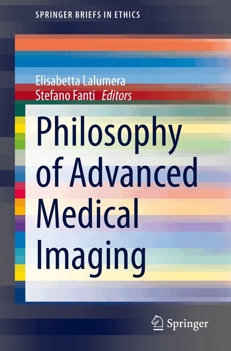
This is the first book to explore the epistemology and ethics of advanced imaging tests, in order to improve the critical understanding of the nature of knowledge they provide and the practical consequences of their utilization in healthcare. Advanced medical imaging tests, such as PET and MRI, …
Philosophy of Advanced Medical Imaging
Av Elisabetta Lalumera, Stefano Fanti
Häftad, 2021
716 kr
Lägg i varukorg
This is the first book to explore the epistemology and ethics of advanced imaging tests, in order to improve the critical understanding of the nature of knowledge they provide and the practical consequences of their utilization in healthcare. Advanced medical imaging tests, such as PET and MRI, …
Atlas of Clinical PET-CT in Treatment Response Evaluation in Oncology
Av Stefano Fanti, Gopinath Gnanasegaran m. fl.
Häftad, 2021
3377 kr
Lägg i varukorg
This atlas is a superb guide to the use of PET-CT for the evaluation of treatment response in oncology patients based on its ability to assess tumor metabolic status. The first part of the book explains the role of PET-CT in response evaluation in different treatment settings. For comparison, …
Atlas of Clinical PET-CT in Treatment Response Evaluation in Oncology
Av Ignasi Carrio, Gopinath Gnanasegaran m. fl.
E-bok, 2021
4187 kr
Lägg i varukorg
This atlas is a superb guide to the use of PET-CT for the evaluation of treatment response in oncology patients based on its ability to assess tumor metabolic status. The first part of the book explains the role of PET-CT in response evaluation in different treatment settings. For comparison, …
Radiology for PET/CT Reporting
Av Cristina Nanni, Stefano Fanti m. fl.
Inbunden, 2022
1355 kr
Lägg i varukorg
This atlas is intended to enable nuclear medicine practitioners who routinely read PET/CT scans to recognize the most common CT abnormalities. Reading PET/CT scans can sometimes be challenging. It is not infrequent, in fact, to encounter abnormal findings in CT images (not related to the neoplastic …
Radiology for PET/CT Reporting
Av Rita Golfieri, Lucia Zanoni m. fl.
E-bok, 2022
1211 kr
Lägg i varukorg
This atlas is intended to enable nuclear medicine practitioners who routinely read PET/CT scans to recognize the most common CT abnormalities. Reading PET/CT scans can sometimes be challenging. It is not infrequent, in fact, to encounter abnormal findings in CT images (not related to the neoplastic …
Radiology for PET/CT Reporting
Av Cristina Nanni, Stefano Fanti m. fl.
Häftad, 2023
962 kr
Lägg i varukorg
This atlas is intended to enable nuclear medicine practitioners who routinely read PET/CT scans to recognize the most common CT abnormalities. Reading PET/CT scans can sometimes be challenging. It is not infrequent, in fact, to encounter abnormal findings in CT images (not related to the neoplastic …
Oncological PET/CT with Histological Confirmation
Av Juliano Cerci, Stefano Fanti m. fl.
Inbunden, 2016
568 kr
Lägg i varukorg
This book reviews the important new field of PET/CT-guided biopsy, which is of potential value in optimizing the diagnostic yield of biopsies. The role of this technology is examined in a range of malignancies, including lymphoma, pancreatic, breast cancer and CNS malignancies. True positive and …
Oncological PET/CT with Histological Confirmation
Av Dominique Delbeke, Stefano Fanti m. fl.
E-bok, 2016
755 kr
Lägg i varukorg
This book reviews the important new field of PET/CT-guided biopsy, which is of potential value in optimizing the diagnostic yield of biopsies. The role of this technology is examined in a range of malignancies, including lymphoma, pancreatic, breast cancer and CNS malignancies. True positive and …
PET/CT in Neuroendocrine Tumors
Av Valentina Ambrosini, Stefano Fanti
Häftad, 2016
848 kr
Lägg i varukorg
This pocket book provides up-to-date descriptions of the most relevant features of neuroendocrine tumors (NETs) and the imaging modalities currently available to assist specialists (clinicians, pathologists, radiologists, nuclear medicine physicians) in selecting optimal patient management based on …
PET/CT in Neuroendocrine Tumors
Av Stefano Fanti, Valentina Ambrosini
E-bok, 2016
1056 kr
Lägg i varukorg
This pocket book provides up-to-date descriptions of the most relevant features of neuroendocrine tumors (NETs) and the imaging modalities currently available to assist specialists (clinicians, pathologists, radiologists, nuclear medicine physicians) in selecting optimal patient management based on …
Oncological PET/CT with Histological Confirmation
Av Juliano Cerci, Stefano Fanti m. fl.
Häftad, 2018
568 kr
Lägg i varukorg
This book reviews the important new field of PET/CT-guided biopsy, which is of potential value in optimizing the diagnostic yield of biopsies. The role of this technology is examined in a range of malignancies, including lymphoma, pancreatic, breast cancer and CNS malignancies. True positive and …
Atlas of PET-CT
A Quick Guide to Image Interpretation
Av Luigi Mansi, Mohsen Farsad m. fl.
E-bok, 2008
755 kr
Lägg i varukorg
This user-friendly atlas, featuring about 500 images, should be a quick guide to interpreting PET/CT images with FDG in oncology. It also illustrates how to recognize normal, para-physiological, and benign pathological uptakes in a case-based practical manner. The text, which includes most relevant …
PET-CT Beyond FDG
A Quick Guide to Image Interpretation
Av Stefano Fanti, Mohsen Farsad m. fl.
Häftad, 2009
1130 kr
Lägg i varukorg
Although [18F]fluorodeoxyglucose (FDG) generally shows an excellent performance as a cancer-imaging agent when using PET-CT, there are some settings in which other radiopharmaceuticals offer advantages. Such non-FDG tracers are now gaining widespread acceptance not only in research but also in …
PET-CT Beyond FDG
A Quick Guide to Image Interpretation
Av Luigi Mansi, Mohsen Farsad m. fl.
E-bok, 2010
1470 kr
Lägg i varukorg
Although [ 18 F]fluorodeoxyglucose (FDG) generally shows an excellent performance as a cancer-imaging agent when using PET-CT, there are some settings in which other radiopharmaceuticals offer advantages. Such non-FDG tracers are now gaining widespread acceptance not only in research but also in …
Atlas of SPECT-CT
Av Stefano Fanti, Mohsen Farsad m. fl.
Häftad, 2011
1130 kr
Lägg i varukorg
The field of nuclear medicine has evolved rapidly in recent years, and one very important aspect of this progress has been the introduction of hybrid imaging systems. PET-CT has already gained widespread acceptance in many clinical settings, especially within oncology, and now SPECT-CT promises to …
Atlas of SPECT-CT
Av Luigi Mansi, Mohsen Farsad m. fl.
E-bok, 2011
1502 kr
Lägg i varukorg
The field of nuclear medicine has evolved rapidly in recent years, and one very important aspect of this progress has been the introduction of hybrid imaging systems. PET-CT has already gained widespread acceptance in many clinical settings, especially within oncology, and now SPECT-CT promises to …
PET-CT: Rare Findings and Diseases
Av Cristina Nanni, Stefano Fanti
Häftad, 2012
568 kr
Lägg i varukorg
PET-CT is increasingly being employed in the diagnosis of both oncological and non-oncological patients, yet nuclear medicine physicians may have only limited practical experience of rare diseases and may experience difficulty in recognizing and interpreting rare findings. This unique atlas …
PET-CT: Rare Findings and Diseases
Av Stefano Fanti, Cristina Nanni
E-bok, 2012
755 kr
Lägg i varukorg
PET-CT is increasingly being employed in the diagnosis of both oncological and non-oncological patients, yet nuclear medicine physicians may have only limited practical experience of rare diseases and may experience difficulty in recognizing and interpreting rare findings. This unique atlas …
Radiology for PET/CT Reporting
Av Lucia Zanoni, Stefano Fanti m. fl.
E-bok, 2013
923 kr
Lägg i varukorg
Reading PET/CT scans is sometimes challenging. Not infrequently, abnormal findings on CT images are functionally silent and therefore difficult for nuclear medicine practitioners to interpret. Furthermore, in general only a low-dose CT scan is produced as part of the combined PET/CT study, and the …
Atlas of PET-CT
A Quick Guide to Image Interpretation
Av Stefano Fanti, Mohsen Farsad m. fl.
Häftad, 2019
1240 kr
Lägg i varukorg
This new atlas, the fourth of a successful series, is a completely revised and updated edition of a previously published FDG PET-CT atlas. In the past few years, considerable progress has been made in the field of PET-CT imaging, and this new edition takes full account of these recent developments. …
Atlas of PET-CT
A Quick Guide to Image Interpretation
Av Paolo Castellucci, Luigi Mansi m. fl.
E-bok, 2019
1670 kr
Lägg i varukorg
This new atlas, the fourth of a successful series, is a completely revised and updated edition of a previously published FDG PET-CT atlas. In the past few years, considerable progress has been made in the field of PET-CT imaging, and this new edition takes full account of these recent developments. …