Luigi Mansi – författare
Visar alla böcker från författaren Luigi Mansi. Handla med fri frakt och snabb leverans.
21 produkter
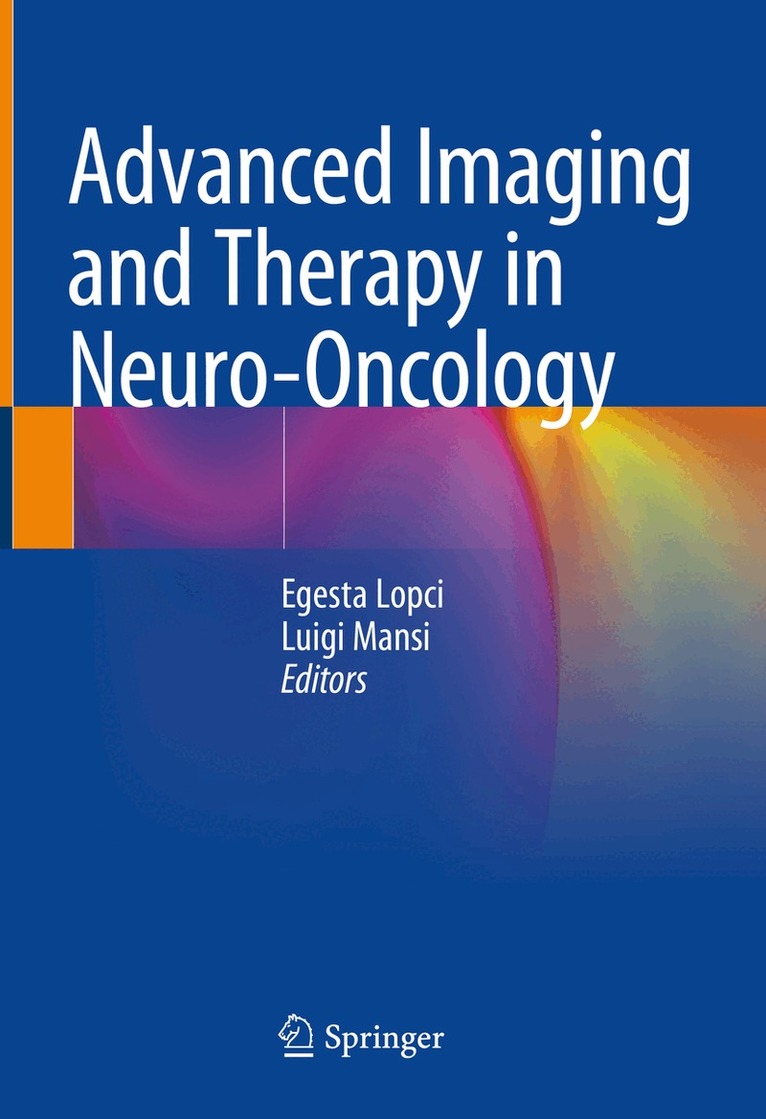
Advanced Imaging and Therapy in Neuro-Oncology
Inbunden, 2024
1576 kr
Lägg i varukorg
This book covers all diagnostic and therapeutic aspects of neuro-oncological diseases. In the last decade, the management of brain tumors has been revolutionized following the new WHO classification and thanks to the introduction of molecular markers into the clinical routine. From an imaging point …
Advanced Imaging and Therapy in Neuro-Oncology
E-bok, 2024
1511 kr
Lägg i varukorg
This book covers all diagnostic and therapeutic aspects of neuro-oncological diseases. In the last decade, the management of brain tumors has been revolutionized following the new WHO classification and thanks to the introduction of molecular markers into the clinical routine. From an imaging point …
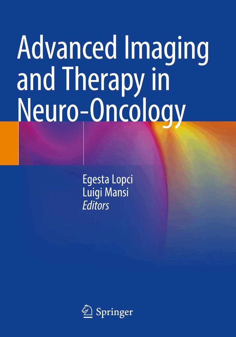
Advanced Imaging and Therapy in Neuro-Oncology
Häftad, 2025
792 kr
Lägg i varukorg
This book covers all diagnostic and therapeutic aspects of neuro-oncological diseases. In the last decade, the management of brain tumors has been revolutionized following the new WHO classification and thanks to the introduction of molecular markers into the clinical routine. From an imaging point …
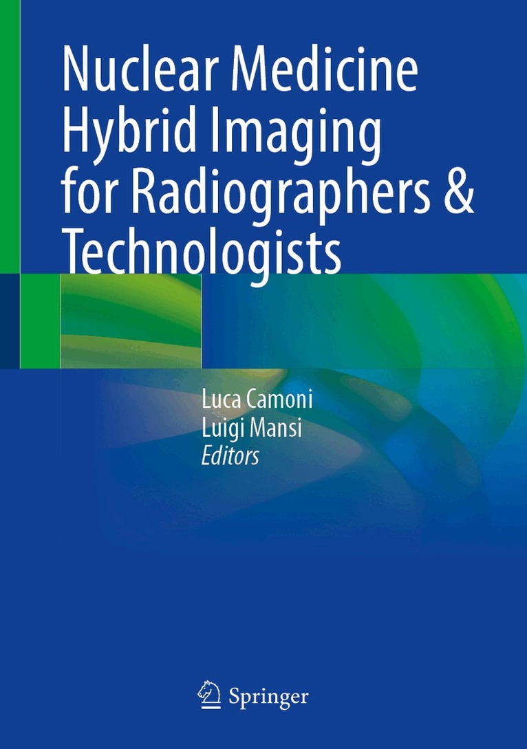
Nuclear Medicine Hybrid Imaging for Radiographers & Technologists
Inbunden, 2025
736 kr
Lägg i varukorg
This essential book, amid the rapid shift toward hybrid imaging, empowers radiographers, technologists and practitioners to build on solid fundamentals, strengthen patient care, and refine advanced techniques.Uniqueness: This book provides a comprehensive perspective on hybrid imaging and patient …
Nuclear Medicine Hybrid Imaging for Radiographers & Technologists
E-bok, 2025
923 kr
Lägg i varukorg
This essential book, amid the rapid shift toward hybrid imaging, empowers radiographers, technologists and practitioners to build on solid fundamentals, strengthen patient care, and refine advanced techniques. Uniqueness: This book provides a comprehensive perspective on hybrid imaging and patient …
Nuclear Medicine Hybrid Imaging for Radiographers & Technologists
Häftad, 2026
568 kr
Lägg i varukorg
This essential book, amid the rapid shift toward hybrid imaging, empowers radiographers, technologists and practitioners to build on solid fundamentals, strengthen patient care, and refine advanced techniques.Uniqueness: This book provides a comprehensive perspective on hybrid imaging and patient …
Clinical Nuclear Medicine in Pediatrics
Av Luigi Mansi, Egesta Lopci m. fl.
Inbunden, 2015
1240 kr
Lägg i varukorg
This book provides a comprehensive state-of-the-art review of pediatric nuclear medicine, encompassing both diagnostic and therapeutic applications. Detailed attention is paid to the role of FDG PET-CT within oncology, but a variety of other long-established or less frequently used diagnostic …
Clinical Nuclear Medicine in Pediatrics
Av Arturo Chiti, Vincenzo Cuccurullo m. fl.
E-bok, 2015
1150 kr
Lägg i varukorg
This book provides a comprehensive state-of-the-art review of pediatric nuclear medicine, encompassing both diagnostic and therapeutic applications. Detailed attention is paid to the role of FDG PET-CT within oncology, but a variety of other long-established or less frequently used diagnostic …
PET-CT and PET-MRI in Neurology
SWOT Analysis Applied to Hybrid Imaging
Av Andrea Ciarmiello, Luigi Mansi
Inbunden, 2016
1352 kr
Lägg i varukorg
Using SWOT analysis, this book examines in detail the strengths and weaknesses of the hybrid modalities PET-CT and PET-MRI for imaging of the central nervous system, comparing their merits and evaluating their advantages over the stand-alone modalities. The aim is to employ a truly systematic …
PET-CT and PET-MRI in Neurology
SWOT Analysis Applied to Hybrid Imaging
Av Luigi Mansi, Andrea Ciarmiello
E-bok, 2016
1224 kr
Lägg i varukorg
Using SWOT analysis, this book examines in detail the strengths and weaknesses of the hybrid modalities PET-CT and PET-MRI for imaging of the central nervous system, comparing their merits and evaluating their advantages over the stand-alone modalities. The aim is to employ a truly systematic …
Clinical Nuclear Medicine in Pediatrics
Av Luigi Mansi, Egesta Lopci m. fl.
Häftad, 2016
904 kr
Lägg i varukorg
This book provides a comprehensive state-of-the-art review of pediatric nuclear medicine, encompassing both diagnostic and therapeutic applications. Detailed attention is paid to the role of FDG PET-CT within oncology, but a variety of other long-established or less frequently used diagnostic …
PET-CT and PET-MRI in Neurology
SWOT Analysis Applied to Hybrid Imaging
Av Andrea Ciarmiello, Luigi Mansi
Häftad, 2018
960 kr
Lägg i varukorg
Using SWOT analysis, this book examines in detail the strengths and weaknesses of the hybrid modalities PET-CT and PET-MRI for imaging of the central nervous system, comparing their merits and evaluating their advantages over the stand-alone modalities. The aim is to employ a truly systematic …
Atlas of PET-CT
A Quick Guide to Image Interpretation
Av Luigi Mansi, Mohsen Farsad m. fl.
E-bok, 2008
755 kr
Lägg i varukorg
This user-friendly atlas, featuring about 500 images, should be a quick guide to interpreting PET/CT images with FDG in oncology. It also illustrates how to recognize normal, para-physiological, and benign pathological uptakes in a case-based practical manner. The text, which includes most relevant …
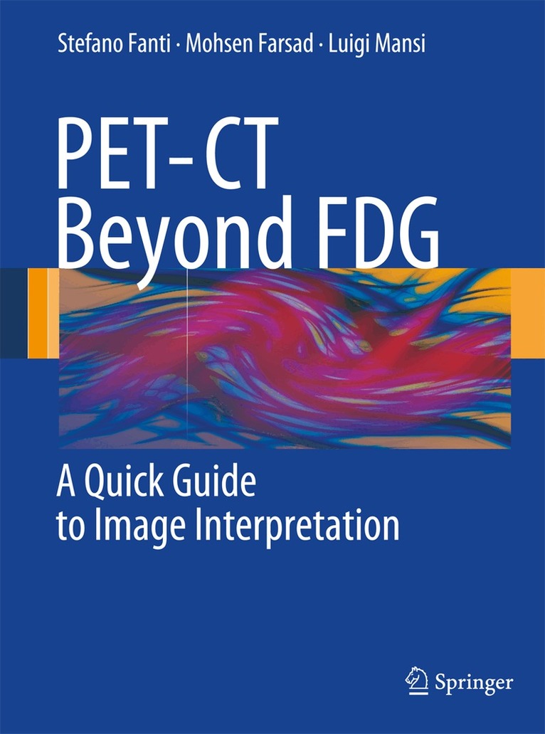
PET-CT Beyond FDG
A Quick Guide to Image Interpretation
Av Stefano Fanti, Mohsen Farsad m. fl.
Häftad, 2009
1128 kr
Lägg i varukorg
Although [18F]fluorodeoxyglucose (FDG) generally shows an excellent performance as a cancer-imaging agent when using PET-CT, there are some settings in which other radiopharmaceuticals offer advantages. Such non-FDG tracers are now gaining widespread acceptance not only in research but also in …
PET-CT Beyond FDG
A Quick Guide to Image Interpretation
Av Luigi Mansi, Mohsen Farsad m. fl.
E-bok, 2010
1470 kr
Lägg i varukorg
Although [ 18 F]fluorodeoxyglucose (FDG) generally shows an excellent performance as a cancer-imaging agent when using PET-CT, there are some settings in which other radiopharmaceuticals offer advantages. Such non-FDG tracers are now gaining widespread acceptance not only in research but also in …
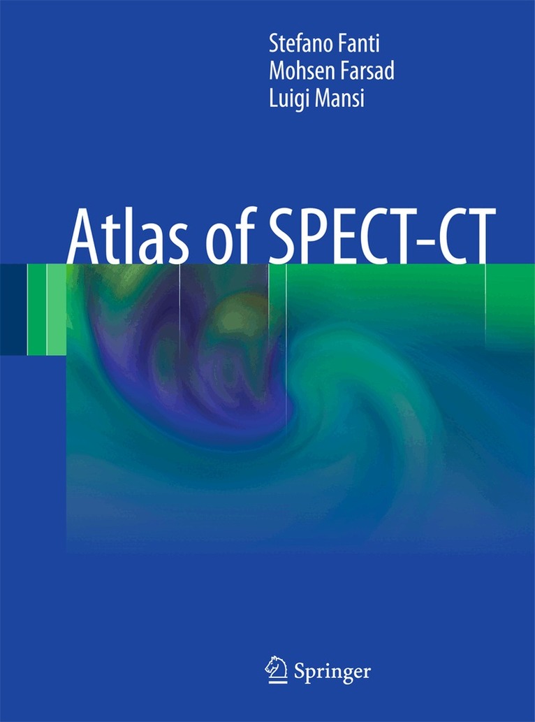
Atlas of SPECT-CT
Av Stefano Fanti, Mohsen Farsad m. fl.
Häftad, 2011
1128 kr
Lägg i varukorg
The field of nuclear medicine has evolved rapidly in recent years, and one very important aspect of this progress has been the introduction of hybrid imaging systems. PET-CT has already gained widespread acceptance in many clinical settings, especially within oncology, and now SPECT-CT promises to …
Atlas of SPECT-CT
Av Luigi Mansi, Mohsen Farsad m. fl.
E-bok, 2011
1502 kr
Lägg i varukorg
The field of nuclear medicine has evolved rapidly in recent years, and one very important aspect of this progress has been the introduction of hybrid imaging systems. PET-CT has already gained widespread acceptance in many clinical settings, especially within oncology, and now SPECT-CT promises to …
Atlas of PET-CT
A Quick Guide to Image Interpretation
Av Stefano Fanti, Mohsen Farsad m. fl.
Häftad, 2019
1240 kr
Lägg i varukorg
This new atlas, the fourth of a successful series, is a completely revised and updated edition of a previously published FDG PET-CT atlas. In the past few years, considerable progress has been made in the field of PET-CT imaging, and this new edition takes full account of these recent developments. …
Atlas of PET-CT
A Quick Guide to Image Interpretation
Av Paolo Castellucci, Luigi Mansi m. fl.
E-bok, 2019
1670 kr
Lägg i varukorg
This new atlas, the fourth of a successful series, is a completely revised and updated edition of a previously published FDG PET-CT atlas. In the past few years, considerable progress has been made in the field of PET-CT imaging, and this new edition takes full account of these recent developments. …