• Fri frakt över 249 kr
  • •
  • Snabba leveranser
  • •
  • Billiga böcker
Kundservice

Du är på sajten för privatpersoner.

Företag, bibliotek eller offentlig verksamhet?

Du handlar på classic.bokus.com, där alla dina funktioner finns intakta.
Till classic.bokus.com
Bokus logotyp. Gå till startsidan.
  • Erbjudanden
  • Nyheter
  • Student
  • Topplistor
  • Barn & ungdom
  • Bokus Play
  • E-böcker
  • Pocketböcker
  • Spel & pussel

10% rabatt på allt med kod NYSTART10 →

Sidfot

Mina sidor

    Hjälp

    • Kundservice
    • Vanliga frågor och svar
    • Frakt och leverans
    • Retur vid ångerrätt
    • Reklamera vara
    • Betalning
    • Köpvillkor
    • Allmänna villkor
    • Information om webbplatsens tillgänglighet

    Om Bokus

    • Om oss
    • Pressrum
    • För studenter
    • För företag
    • För bibliotek och offentlig verksamhet
    • För leverantörer
    • Hållbarhet

    Populärt

    • Aktuella erbjudanden
    • Presentkort
    • Studentlitteratur
    • Nya böcker
    • Topplistor
    • Signerade böcker
    • Engelska böcker

    Inspiration

    • Boktips
    • BookTok
    • Populära bokserier
    • Barnbokskaraktärer
    • Populära författare
    Logotyp för Bokus
    Följ oss på Facebook (extern länk)Följ oss på Instagram (extern länk)Följ oss på YouTube (extern länk)Följ oss på TikTok (extern länk)
    bokus @ CookiesAnpassa cookiesIntegritetspolicyKöpvillkor
    Till Citymail hemsida (extern länk)Till Budbee hemsida (extern länk)Till Postnord hemsida (extern länk)Till Schenker hemsida (extern länk)Till Early Bird hemsida (extern länk)Till Walleys hemsida (extern länk)
    1. Medicin
    2. Andra medicinska specialiteter
    3. Medicinsk bildbehandling

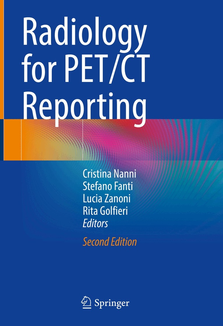
    Radiology for PET/CT Reporting

    AvCristina Nanni,Stefano Fanti

    Inbunden, Engelska, 2022

    1 339 kr

    Beställningsvara. Skickas inom 10-15 vardagar. Fri frakt över 249 kr.

    Beskrivning

    This atlas is intended to enable nuclear medicine practitioners who routinely read PET/CT scans to recognize the most common CT abnormalities. Reading PET/CT scans can sometimes be challenging. It is not infrequent, in fact, to encounter abnormal findings in CT images (not related to the neoplastic disease under evaluation) that are functionally silent and therefore difficult to interpret for nuclear medicine practitioners. Frequently, these findings are clinically relevant and should be reported, interpreted and compared to previous scans. This may also have an impact on patient management, since expensive tests like PET/CT are expected to provide the highest level of diagnostic information. Generally, CT images associated with a PET scan are acquired in a low-dose modality, and therefore prove to be sub-optimal for CT image interpretation. Sometimes a comparison with a full-resolution and contrast-enhanced CT atlas may be difficult. Low-dose CT slices are thicker than diagnostic CT and offer less anatomical detail, which can affect accuracy in terms of recognizing both anatomical structures and pathological findings.Today it is becoming increasingly common to acquire a standard PET/CT by combining the administration of contrast media and a diagnostic CT; here, too, basic CT reporting skills are needed in clinical practice. This atlas features a chapter on “normal anatomy” (with and without contrast media) that is based on low-dose and full-dose CT images from PET/CT standard acquisition, and which identifies all the relevant anatomical structures. Other chapters (focusing on the thorax, abdomen, pelvis, and musculoskeletal system) present cases with common and uncommon anatomical abnormalities. The addition of new cases with ceCT in this revised second edition rounds out the coverage of PET/CT reporting. Given its scope, the book will be of interest to nuclear medicine physicians, radiologists, and oncologists alike.

    Produktinformation

    • Utgivningsdatum:2022-04-14
    • Mått:178 x 254 x 18 mm
    • Vikt:710 g
    • Format:Inbunden
    • Språk:Engelska
    • Antal sidor:229
    • Upplaga:2
    • Förlag:Springer Nature Switzerland AG
    • ISBN:9783030876401
    • Originaltitel:Radiology for PET/CT Reporting

    Utforska kategorier

    • Medicinsk bildbehandling inom Medicin

    Mer om författaren

    Dr. Cristina Nanni is a Nuclear Medicine Physician who has been working at the PET Centre in Bologna, Italy, since 2007. She completed her residency in Nuclear Medicine at the University of Bologna (2004) and in Radiology in 2012. She worked in the field of pre-clinical PET imaging in 2005 and 2006, and is currently responsible for new radiotracer applications and the integration of morphological and functional imaging (PET/ceCT and PET/CT guided biopsy) into clinical practice. She is the author or co-author of more than 200 clinical and pre-clinical papers published in international journals, and of more than 200 abstracts presented at national and international congresses.Dr. Lucia Zanoni is a Nuclear Medicine Physician who has been working at the PET Centre in Bologna, Italy, since 2016. She completed her residency in Nuclear Medicine at the University of Bologna (2015). Chiefly active in the field of oncological PET/CT, she is the author or co-author of 30 clinical and pre-clinical papers published in international journals.Prof. Stefano Fanti is the Head of Nuclear Medicine at AOU S. Orsola Malpighi and a Professor of Nuclear Medicine at the University of Bologna. He is the author of more than 470 publications.Prof. Rita Golfieri is the Head of Radiology at AOU S. Orsola Malpighi and a Professor of Radiology at the University of Bologna. She is the author of 10 books, 70 book chapters, and nearly 300 articles published in respected national and international journals.

    Innehållsförteckning

    • Chapter 1: Normal Anatomy.- Chapter 2:  Head & Neck.- Chapter 3: Thorax.- Chapter 4: Abdomen.-  Chapter 5: Pelvis.- Chapter 6:  Musculoskeletal.