V. Chan-Palay – författare
Visar alla böcker från författaren V. Chan-Palay. Handla med fri frakt och snabb leverans.
5 produkter
5 produkter
E-bok
PDF, Engelska, 20121 947 kr
Läs direkt efter köp
The origins of this book go back to the first electron microscopic studies of the central nervous system. The cerebellar cortex was from the first an object of close study in the electron microscope, repeating in modern cytology and neuroanatomy the role it had in the hands of RAMON y CAJAL at the end of the nineteenth century. The senior author vividly remembers a day early in 1953 when GEORGE PALADE, with whom he was then working, showed him an electron micrograph of a cerebellar glomerulus, saying "That is what the synapse should look like. " It is true that the tissue was swollen and the mitochondria were exploded, but all of the essentials of synaptic structure were visible. At that time small fragments of tissue, fixed by immersion in osmium tetroxide and embedded in methacrylate, were laboriously sectioned with glass knives without any predetermined orientation and then examined in the electron microscope. After much searching, favorably preserved areas'' were studied at the cytological level in order to recognize the parts of neurons and characterize them. Such procedures, dependent upon random sections and uncontrollable selection by a highly erratic technique of preservation, precluded any systematic investigation of the organization of a particular nucleus or region of the central nervous system. It was difficult enough to distinguish neurons from the neuroglia.
Häftad, Engelska, 2011
1 560 kr
Skickas inom 10-15 vardagar
The origins of this book go back to the first electron microscopic studies of the central nervous system. The cerebellar cortex was from the first an object of close study in the electron microscope, repeating in modern cytology and neuroanatomy the role it had in the hands of RAMON y CAJAL at the end of the nineteenth century. The senior author vividly remembers a day early in 1953 when GEORGE PALADE, with whom he was then working, showed him an electron micrograph of a cerebellar glomerulus, saying "That is what the synapse should look like. " It is true that the tissue was swollen and the mitochondria were exploded, but all of the essentials of synaptic structure were visible. At that time small fragments of tissue, fixed by immersion in osmium tetroxide and embedded in methacrylate, were laboriously sectioned with glass knives without any predetermined orientation and then examined in the electron microscope. After much searching, favorably preserved areas' were studied at the cytological level in order to recognize the parts of neurons and characterize them. Such procedures, dependent upon random sections and uncontrollable selection by a highly erratic technique of preservation, precluded any systematic investigation of the organization of a particular nucleus or region of the central nervous system. It was difficult enough to distinguish neurons from the neuroglia.
E-bok
PDF, Engelska, 20131 785 kr
Läs direkt efter köp
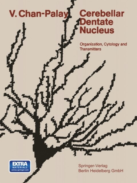
Recent physiologic investigations have shown that the deep cerebellar nuclei may play an important role in the initiation and monitoring of skilled move ments. Much of this physiologic work has been carried out in the absence of a secure foundation in neuroanatomical information. Although the main sources of the afferent fibers and the major terminations of the efferent fibers related to these nuclei have been known for many years, remarkably little information about the organization of the nuclei themselves has been collected. The kinds of nerve cells, their arrangement within the nuclei, the patterns of their dendritic arborizations, the distribution of incoming fibers among the neurons, the relationship between the outgoing nerve fibers and the nerve cells from which they originate - these and many other morphologic features were either unknown or only superficially explored. In fact, so little was known about the deep cerebellar nuclei when I began to work on this subject that the investigations reported here are virtually without antecedents, a refreshing change from the cerebellar cortex which has been repeatedly and exhaustively surveyed. My studies on the cerebellar nuclei began in the spring of 1972. They were initiated with the intent of applying the principles of analysis that had been developed for the cerebellar cortex to a different but related part of the brain.
Häftad, Engelska, 2014
1 338 kr
Skickas inom 10-15 vardagar
Recent physiologic investigations have shown that the deep cerebellar nuclei may play an important role in the initiation and monitoring of skilled move ments. Much of this physiologic work has been carried out in the absence of a secure foundation in neuroanatomical information. Although the main sources of the afferent fibers and the major terminations of the efferent fibers related to these nuclei have been known for many years, remarkably little information about the organization of the nuclei themselves has been collected. The kinds of nerve cells, their arrangement within the nuclei, the patterns of their dendritic arborizations, the distribution of incoming fibers among the neurons, the relationship between the outgoing nerve fibers and the nerve cells from which they originate - these and many other morphologic features were either unknown or only superficially explored. In fact, so little was known about the deep cerebellar nuclei when I began to work on this subject that the investigations reported here are virtually without antecedents, a refreshing change from the cerebellar cortex which has been repeatedly and exhaustively surveyed. My studies on the cerebellar nuclei began in the spring of 1972. They were initiated with the intent of applying the principles of analysis that had been developed for the cerebellar cortex to a different but related part of the brain.
Häftad, Engelska, 2011
1 671 kr
Skickas inom 10-15 vardagar
The present volume consists of papers prepared for a conference on the cerebellum which was held at the National Institutes of Health in Bethesda on 15 -17 May 1980. This was the first gen eral conference on the cerebellum since the 1972 symposium in Portland, Oregon, which was convened to celebrate the publica tion of the last volume of the Comparative Anatomy and Histolo gy of the Cerebellum by Larsell and Jansen. In organizing the 1980 Conference, we elected to emphasize ad vances in neuroanatomy over other aspects because, in our view, morphological investigation continues to playa large and essential role in the developing understanding of cerebellar func tion, and, despite the general impression that cerebellar anat omy is better known than any other part of the nervous system, our information on this topic is far from complete. N everthe less, the cerebellum offers the best model we have for analyzing the vertebrate central nervous system. The correlation of ana tomical, cytological, developmental, physiological, chemical, and pharmacological data on this relatively simple and uniform structure, which has persisted with remarkable conservatism through vertebrate evolution, still holds a rich store of answers to the most penetrating questions concerning the functional organization of the brain. Naturally, within the limits of time, space, and our resources we could not pretend to include every thing that has been learned about the cerebellum in the past decade.