
RMS - Royal Microscopical Society – serie
Visar alla böcker i serien RMS - Royal Microscopical Society. Handla med fri frakt och snabb leverans.
9 produkter
9 produkter
Inbunden, Engelska, 2008
970 kr
Skickas inom 5-8 vardagar
Offers a simple starting point to VPSEM, especially for new users, technicians and students containing clear, concise explanationsCrucially, the principles and applications outlined in this book are completely generic: i.e. applicable to all types of VPSEM, irrespective of manufacturer.Information presented will enable reader to turn principles into practicePublished in association with the Royal Microscopical Society (RMS) -www.rms.org.uk
Inbunden, Engelska, 2011
762 kr
Skickas inom 5-8 vardagar
The book is concerned with the theory, background, and practical use of transmission electron microscopes with lens correctors that can correct the effects of spherical aberration. The book also covers a comparison with aberration correction in the TEM and applications of analytical aberration corrected STEM in materials science and biology. This book is essential for microscopists involved in nanoscale and materials microanalysis especially those using scanning transmission electron microscopy, and related analytical techniques such as electron diffraction x-ray spectrometry (EDXS) and electron energy loss spectroscopy (EELS).
Inbunden, Engelska, 2019
2 202 kr
Skickas inom 5-8 vardagar
Introduces readers to the enlightening world of the modern light microscopeThere have been rapid advances in science and technology over the last decade, and the light microscope, together with the information that it gives about the image, has changed too. Yet the fundamental principles of setting up and using a microscope rests upon unchanging physical principles that have been understood for years. This informative, practical, full-colour guide fills the gap between specialised edited texts on detailed research topics, and introductory books, which concentrate on an optical approach to the light microscope. It also provides comprehensive coverage of confocal microscopy, which has revolutionised light microscopy over the last few decades. Written to help the reader understand, set up, and use the often very expensive and complex modern research light microscope properly, Understanding Light Microscopy keeps mathematical formulae to a minimum—containing and explaining them within boxes in the text. Chapters provide in-depth coverage of basic microscope optics and design; ergonomics; illumination; diffraction and image formation; reflected-light, polarised-light, and fluorescence microscopy; deconvolution; TIRF microscopy; FRAP & FRET; super-resolution techniques; biological and materials specimen preparation; and more. Gives a didactic introduction to the light microscopeEncourages readers to use advanced fluorescence and confocal microscopes within a research institute or core microscopy facilityFeatures full-colour illustrations and workable practical protocolsUnderstanding Light Microscopy is intended for any scientist who wishes to understand and use a modern light microscope. It is also ideal as supporting material for a formal taught course, or for individual students to learn the key aspects of light microscopy through their own study.
Inbunden, Engelska, 2018
1 466 kr
Skickas inom 5-8 vardagar
A detailed presentation of the physics of electron beam-specimen interactionsElectron microscopy is one of the most widely used characterisation techniques in materials science, physics, chemistry, and the life sciences. This book examines the interactions between the electron beam and the specimen, the fundamental starting point for all electron microscopy. Detailed explanations are provided to help reinforce understanding, and new topics at the forefront of current research are presented. It provides readers with a deeper knowledge of the subject, particularly if they intend to simulate electron beam-specimen interactions as part of their research projects. The book covers the vast majority of commonly used electron microscopy techniques. Some of the more advanced topics (annular bright field and dopant atom imaging, atomic resolution chemical analysis, band gap measurements) provide additional value, especially for readers who have access to advanced instrumentation, such as aberration-corrected and monochromated microscopes.Electron Beam-Specimen Interactions and Simulation Methods in Microscopy offers enlightening coverage of: the Monte-Carlo Method; Multislice Simulations; Bloch Waves in Conventional and Analytical Transmission Electron Microscopy; Bloch Waves in Scanning Transmission Electron Microscopy; Low Energy Loss and Core Loss EELS. It also supplements each chapter with clear diagrams and provides appendices at the end of the book to assist with the pre-requisites. A detailed presentation of the physics of electron beam-specimen interactionsEach chapter first discusses the background physics before moving onto simulation methodsUses computer programs to simulate electron beam-specimen interactions (presented in the form of case studies)Includes hot topics brought to light due to advances in instrumentation (particularly aberration-corrected and monochromated microscopes)Electron Beam-Specimen Interactions and Simulation Methods in Microscopy benefits students undertaking higher education degrees, practicing electron microscopists who wish to learn more about their subject, and researchers who wish to obtain a deeper understanding of the subject matter for their own work.
Inbunden, Engelska, 2019
2 195 kr
Skickas inom 5-8 vardagar
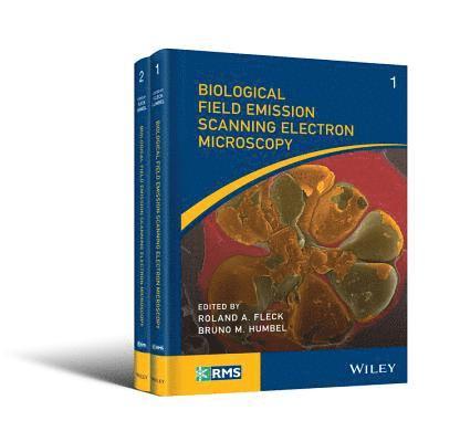
The go‐to resource for microscopists on biological applications of field emission gun scanning electron microscopy (FEGSEM)The evolution of scanning electron microscopy technologies and capability over the past few years has revolutionized the biological imaging capabilities of the microscope—giving it the capability to examine surface structures of cellular membranes to reveal the organization of individual proteins across a membrane bilayer and the arrangement of cell cytoskeleton at a nm scale. Most notable are their improvements for field emission scanning electron microscopy (FEGSEM), which when combined with cryo-preparation techniques, has provided insight into a wide range of biological questions including the functionality of bacteria and viruses. This full-colour, must-have book for microscopists traces the development of the biological field emission scanning electron microscopy (FEGSEM) and highlights its current value in biological research as well as its future worth. Biological Field Emission Scanning Electron Microscopy highlights the present capability of the technique and informs the wider biological science community of its application in basic biological research. Starting with the theory and history of FEGSEM, the book offers chapters covering: operation (strengths and weakness, sample selection, handling, limitations, and preparation); Commercial developments and principals from the major FEGSEM manufacturers (Thermo Scientific, JEOL, HITACHI, ZEISS, Tescan); technical developments essential to bioFEGSEM; cryobio FEGSEM; cryo-FIB; FEGSEM digital-tomography; array tomography; public health research; mammalian cells and tissues; digital challenges (image collection, storage, and automated data analysis); and more. Examines the creation of the biological field emission gun scanning electron microscopy (FEGSEM) and discusses its benefits to the biological research community and future valueProvides insight into the design and development philosophy behind current instrument manufacturersCovers sample handling, applications, and key supporting techniquesFocuses on the biological applications of field emission gun scanning electron microscopy (FEGSEM), covering both plant and animal researchPresented in full colourAn important part of the Wiley-Royal Microscopical Series, Biological Field Emission Scanning Electron Microscopy is an ideal general resource for experienced academic and industrial users of electron microscopy—specifically, those with a need to understand the application, limitations, and strengths of FEGSEM.
Inbunden, Engelska, 2019
1 538 kr
Skickas inom 5-8 vardagar
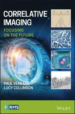
Brings a fresh point of view to the current state of correlative imaging and the future of the fieldThis book provides contributions from international experts on correlative imaging, describing their vision of future developments in the field based on where it is today. Starting with a brief historical overview of how the field evolved, it presents the latest developments in microscopy that facilitate the correlative workflow. It also discusses the need for an ideal correlative probe, applications in proteomic and elemental analysis, interpretation methods, and how correlative imaging can incorporate force microscopy, soft x-ray tomography, and volume electron microscopy techniques. Work on placing individual molecules within cells is also featured.Correlative Imaging: Focusing on the Future offers in-depth chapters on: correlative imaging from an LM perspective; the importance of sample processing for correlative imaging; correlative light and volume EM; correlation with scanning probe microscopies; and integrated microscopy. It looks at: cryo-correlative microscopy; correlative cryo soft X-ray imaging; and array tomography. Hydrated-state correlative imaging in vacuo, correlating data from different imaging modalities, and big data in correlative imaging are also considered. Brings a fresh view to one of the hottest topics within the imaging community: the correlative imaging fieldDiscusses current research and offers expert thoughts on the field’s future developmentsPresented by internationally-recognized editors and contributors with extensive experience in research and applicationsOf interest to scientists working in the fields of imaging, structural biology, cell biology, developmental biology, neurobiology, cancer biology, infection and immunity, biomaterials and biomedicinePart of the Wiley–Royal Microscopical Society seriesCorrelative Imaging: Focusing on the Future will appeal to those working in the expanding field of the biosciences, correlative microscopy and related microscopic areas. It will also benefit graduate students working in microscopy, as well as anyone working in the microscopy imaging field in biomedical research.
Inbunden, Engelska, 2013
1 294 kr
Skickas
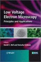
Part of the Wiley-Royal Microscopical Society Series, this book discusses the rapidly developing cutting-edge field of low-voltage microscopy, a field that has only recently emerged due to the rapid developments in the electron optics design and image processing.It serves as a guide for current and new microscopists and materials scientists who are active in the field of nanotechnology, and presents applications in nanotechnology and research of surface-related phenomena, allowing researches to observe materials as never before.
Inbunden, Engelska, 2012
1 268 kr
Skickas inom 5-8 vardagar
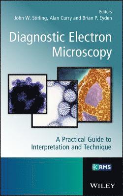
Diagnostic Electron MicroscopyDiagnostic Electron Microscopy: A Practical Guide to Interpretation and Technique summarises the current interpretational applications of TEM in diagnostic pathology. This concise and accessible volume provides a working guide to the main, or most useful, applications of the technique including practical topics of concern to laboratory scientists, brief guides to traditional tissue and microbiological preparation techniques, microwave processing, digital imaging and measurement uncertainty.The text features both a screening and interpretational guide for TEM diagnostic applications and current TEM diagnostic tissue preparation methods pertinent to all clinical electron microscope units worldwide. Containing high-quality representative images, this up-to-date text includes detailed information on the most important diagnostic applications of transmission electron microscopy as well as instructions for specific tissues and current basic preparative techniques.The book is relevant to trainee pathologists and practising pathologists who are expected to understand and evaluate/screen tissues by TEM. In addition, technical and scientific staff involved in tissue preparation and diagnostic tissue evaluation/screening by TEM will find this text useful.
Inbunden, Engelska, 2026
1 631 kr
Skickas inom 5-8 vardagar
COMPREHENSIVE REFERENCE EXPLORING MICROSCOPY TECHNIQUES TO SUPPORT THE STUDY OF LIVING SYSTEMS WITH A UNIQUE MULTISCALE FOCUS Live Biological Imaging Across Scales presents an overview of the technologies and experimental strategies available for live imaging across scales. Each chapter highlights a different technique, working upwards from small to large scale. Readers will find critical evaluation of these techniques and perspectives on future developments in this evolving field, enabling them to effectively identify techniques that may benefit their research activities. Written by a team of leading experts and part of the Wiley-Royal Microscopical Society series, Live Biological Imaging Across Scales includes information on: Single cells, covering single molecule tracking/FRET, actin, microtubules, intermediate filaments, septins, vesicles, phagosomes, and confined environmentsMultiple cells, covering collective migration, endothelial monolayers, flow platelets, and flow, wound healing, monocyte transmigration, cancer spheroids/tumor models, and organoidsOrganisms, covering Dictyostelium, C. elegans, Drosophila, and mouse-tumorsImaging of whole cells individually and collectively in both 2D and 3D platformsLive Biological Imaging Across Scales is an essential reference for microscopists and experimental biologists, particularly those interested and/or working in the expanding field of live imaging in biology, as well as those working in the commercial microscopy industry supply and development.