
Series in Cellular and Clinical Imaging – serie
Visar alla böcker i serien Series in Cellular and Clinical Imaging. Handla med fri frakt och snabb leverans.
16 produkter
16 produkter

Häftad, Engelska, 2023
746 kr
Skickas inom 10-15 vardagar
Imaging from Cells to Animals In Vivo offers an overview of optical imaging techniques developed over the past two decades to investigate biological processes in live cells and tissues. It comprehensively covers the main imaging approaches used as well as the application of those techniques to biological investigations in preclinical models. Among the areas covered are cell metabolism, receptor-ligand interactions, membrane trafficking, cell signaling, cell migration, cell adhesion, cytoskeleton and other processes using various molecular optical imaging techniques in living organisms, such as mice and zebrafish.FeaturesBrings together biology and advanced optical imaging techniques to provide an overview of progress and modern methods from microscopy to whole body imaging.Fills the need for a comprehensive view of application-driven development and use of new tools to ask new biological questions in the context of a living system.Includes basic chapters on key methods and instrumentation, from fluorescence microscopy and imaging to endoscopy, optical coherence tomography and super-resolution imaging.Discusses approaches at different length scales and biomedical applications to the study of single cell, whole organ, and whole organism behavior.Addresses the impact on discovery, such as cellular function as implicated in human disease and translational medicine, for example in cancer diagnosis.
Häftad, Engelska, 2021
790 kr
Skickas inom 10-15 vardagar
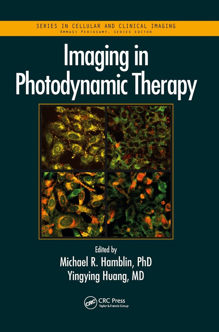
This book covers the broad field of cellular, molecular, preclinical, and clinical imaging either associated with or combined with photodynamic therapy (PDT). It showcases how this approach is used clinically for cancer, infections, and diseases characterized by unwanted tissue such as atherosclerosis or blindness. Because the photosensitizers are also fluorescent, the book also addresses various imaging systems such as confocal microscopy and small animal imaging systems, and highlights how they have been used to follow and optimize treatment, and to answer important mechanistic questions. Chapters also discuss how imaging has made important contributions to clinical outcomes in skin, bladder, and brain cancers, as well as in the development of theranostic agents for detection and treatment of disease. This book provides a resource for physicians and research scientists in cell biology, microscopy, optics, molecular imaging, oncology, and drug discovery.
Häftad, Engelska, 2021
775 kr
Skickas inom 10-15 vardagar
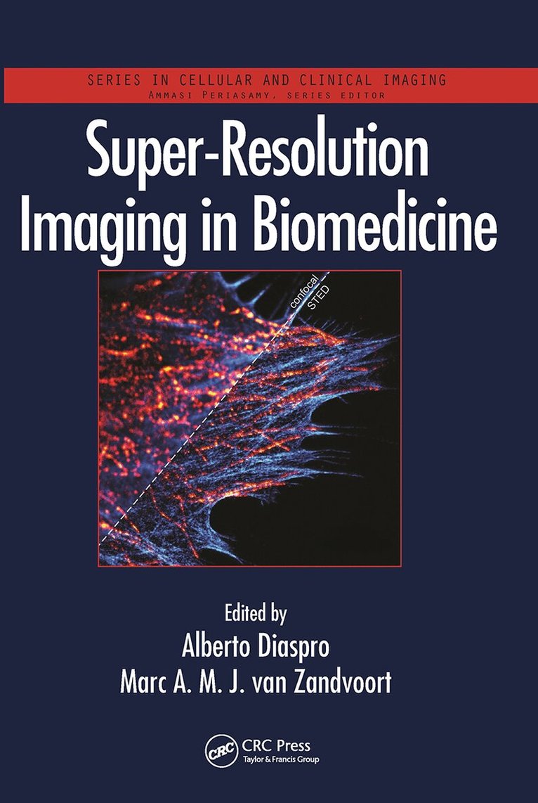
This book encompasses the full breadth of the super-resolution imaging field, representing modern techniques that exceed the traditional diffraction limit, thereby opening up new applications in biomedicine. It shows readers how to use the new tools to increase resolution in sub-nanometer-scale images of living cells and tissue, which leads to new information about molecules, pathways and dynamics. The book highlights the advantages and disadvantages of the techniques, and gives state-of-the-art examples of applications using microscopes currently available on the market. It covers key techniques such as stimulated emission depletion (STED), structured illumination microscopy (SSIM), photoactivated localization microscopy (PALM), and stochastic optical reconstruction microscopy (STORM). It will be a useful reference for biomedical researchers who want to work with super-resolution imaging, learn the proper technique for their application, and simultaneously obtain a solid footing in other techniques.
Inbunden, Engelska, 2023
1 819 kr
Skickas inom 10-15 vardagar
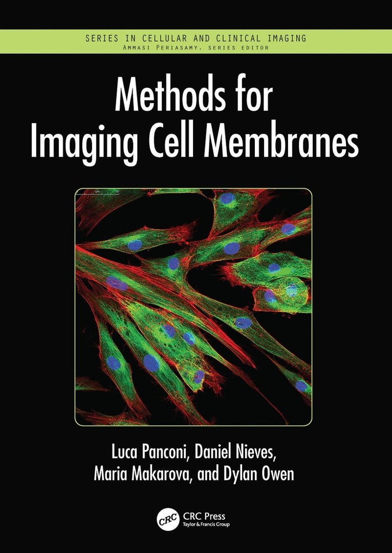
This book will serve as an introduction to microscopy and biomedical imaging methods, with a focus on the study of the distributions and dynamics of molecules on the cell surface. It will provide readers with an in-depth understanding of how modern microscopy methodology can be used to understand the organisation of cell membrane systems and how experiments can be designed around these methodologies.There are numerous methods employed to understand cell membrane organisation, but foremost among them are microscopy methods which can map the distributions of molecules on the cell surface and even map the biophysical properties of membranes themselves. Fluorescence microscopy has been especially widely used due to its specificity and relatively noninvasive nature, allowing live-cell imaging. However, the recent advance of super-resolution fluorescence microscopy has broken the previous resolution limit for this type of microscopy, which has been an important advancement in the field. Atomic force microscopy and electron microscopy have also been deployed to learn about membrane organisation and properties.Each chapter in this volume will be themed around measuring a particular property of cell membranes. In each case, the authors examine the range of methodology applicable to the task, comparing the advantages and disadvantages of each one, and will also provide an overview of important discoveries that have been made using the methodology being discussed. The chapters will cover:Measuring membrane protein distributions using single-molecule localisation microscopy (SMLM)Measuring membrane protein dynamics and diffusion using fluorescence correla-tion spectroscopy (FCS)Mapping membrane lipid backing using environmentally sensitive fluorescence probesMapping membrane thickness and rigidity using atomic force microscopyMapping membrane proteins and the cytoskeleton using electron microscopyThis book will be a valuable resource to graduate and upper-level undergraduate students and industry researchers in the fields of cell biology, microbiology, microscopy, and medical imaging.
Häftad, Engelska, 2025
795 kr
Skickas inom 10-15 vardagar
This book will serve as an introduction to microscopy and biomedical imaging methods, with a focus on the study of the distributions and dynamics of molecules on the cell surface. It will provide readers with an in-depth understanding of how modern microscopy methodology can be used to understand the organisation of cell membrane systems and how experiments can be designed around these methodologies.There are numerous methods employed to understand cell membrane organisation, but foremost among them are microscopy methods which can map the distributions of molecules on the cell surface and even map the biophysical properties of membranes themselves. Fluorescence microscopy has been especially widely used due to its specificity and relatively noninvasive nature, allowing live-cell imaging. However, the recent advance of super-resolution fluorescence microscopy has broken the previous resolution limit for this type of microscopy, which has been an important advancement in the field. Atomic force microscopy and electron microscopy have also been deployed to learn about membrane organisation and properties.Each chapter in this volume will be themed around measuring a particular property of cell membranes. In each case, the authors examine the range of methodology applicable to the task, comparing the advantages and disadvantages of each one, and will also provide an overview of important discoveries that have been made using the methodology being discussed. The chapters will cover:Measuring membrane protein distributions using single-molecule localisation microscopy (SMLM)Measuring membrane protein dynamics and diffusion using fluorescence correla-tion spectroscopy (FCS)Mapping membrane lipid backing using environmentally sensitive fluorescence probesMapping membrane thickness and rigidity using atomic force microscopyMapping membrane proteins and the cytoskeleton using electron microscopyThis book will be a valuable resource to graduate and upper-level undergraduate students and industry researchers in the fields of cell biology, microbiology, microscopy, and medical imaging.
Häftad, Engelska, 2022
790 kr
Skickas inom 10-15 vardagar
The Handbook of Neurophotonics provides a dedicated overview of neurophotonics, covering the use of advanced optical technologies to record, stimulate, and control the activity of the brain, yielding new insight and advantages over conventional tools due to the adaptability and non-invasive nature of light.Including 30 colour figures, this book addresses functional studies of neurovascular signaling, metabolism, electrical excitation, and hemodynamics, as well as clinical applications for imaging and manipulating brain structure and function. The unifying theme throughout is not only to highlight the technology, but to show how these novel methods are becoming critical to breakthroughs that will lead to advances in our ability to manage and treat human diseases of the brain.Key Features:Provides the first dedicated book on state-of-the-art optical techniques for sensing and imaging across at the cellular, molecular, network, and whole brain levels.Highlights how the methods are used for measurement, control, and tracking of molecular events in live neuronal cells, both in basic research and clinical practice.Covers the entire spectrum of approaches, from optogenetics to functional methods, photostimulation, optical dissection, multiscale imaging, microscopy, and structural imaging.Includes chapters that show use of voltage-sensitive dye imaging, hemodynamic imaging, multiphoton imaging, temporal multiplexing, multiplane microscopy, optoacoustic imaging, near-infrared spectroscopy, and miniature neuroimaging devices to track cortical brain activity.
Inbunden, Engelska, 2020
3 052 kr
Skickas inom 10-15 vardagar
Imaging from Cells to Animals In Vivo offers an overview of optical imaging techniques developed over the past two decades to investigate biological processes in live cells and tissues. It comprehensively covers the main imaging approaches used as well as the application of those techniques to biological investigations in preclinical models. Among the areas covered are cell metabolism, receptor-ligand interactions, membrane trafficking, cell signaling, cell migration, cell adhesion, cytoskeleton and other processes using various molecular optical imaging techniques in living organisms, such as mice and zebrafish.FeaturesBrings together biology and advanced optical imaging techniques to provide an overview of progress and modern methods from microscopy to whole body imaging.Fills the need for a comprehensive view of application-driven development and use of new tools to ask new biological questions in the context of a living system.Includes basic chapters on key methods and instrumentation, from fluorescence microscopy and imaging to endoscopy, optical coherence tomography and super-resolution imaging.Discusses approaches at different length scales and biomedical applications to the study of single cell, whole organ, and whole organism behavior.Addresses the impact on discovery, such as cellular function as implicated in human disease and translational medicine, for example in cancer diagnosis.
Häftad, Engelska, 2018
1 007 kr
Skickas inom 10-15 vardagar
From the Lab to Clinical Settings—Advances in Quantitative, Noninvasive Optical DiagnosticsNoninvasive fluorescence imaging techniques, novel fluorescent labels, and natural biomarkers are revolutionizing our knowledge of cellular processes, signaling and metabolic pathways, the underlying mechanisms for health problems, and the identification of new therapeutic targets for drug discoveries. Natural Biomarkers for Cellular Metabolism: Biology, Techniques, and Applications delves into the current state of knowledge on intrinsic fluorescent biomarkers and highlights recent developments in using these biomarkers for the metabolic mapping and clinical diagnosis of healthy and diseased cells and tissues.Autofluorescent Biomarkers for Biomedical DiagnosticsThe book’s first section introduces the fundamentals of cellular energy metabolism as well as natural biomarkers within the context of their biological functions. The second section outlines the theoretical and technical background of quantitative, noninvasive, autofluorescence microscopy and spectroscopy methods, including experimental design, calibration, pitfalls, and remedies of data acquisition and analysis. The last two sections highlight advances in biomedical and biochemical applications, such as monitoring stem cell differentiation in engineered tissues and diagnosing cancer and ophthalmic diseases quantitatively and noninvasively.Tailored to Interdisciplinary Researchers Covering cell biology, imaging techniques, and clinical diagnostics, this book provides readers with a complete guide to studying cellular/tissue metabolism under healthy, diseased, and environment-induced stress conditions using natural biomarkers. The book is designed for graduate and advanced undergraduate students, biophysics instructors, medical researchers, and those in pharmaceutical R&D.
Häftad, Engelska, 2018
1 210 kr
Skickas inom 10-15 vardagar
The First Book on CRS MicroscopyCompared to conventional Raman microscopy, coherent Raman scattering (CRS) allows label-free imaging of living cells and tissues at video rate by enhancing the weak Raman signal through nonlinear excitation. Edited by pioneers in the field and with contributions from a distinguished team of experts, Coherent Raman Scattering Microscopy explains how CRS can be used to obtain a point-by-point chemical map of live cells and tissues.In color throughout, the book starts by establishing the foundation of CRS microscopy. It discusses the principles of nonlinear optical spectroscopy, particularly coherent Raman spectroscopy, and presents the theories of contrast mechanisms pertinent to CRS microscopy. The text then provides important technical aspects of CRS microscopy, including microscope construction, detection schemes, and data analyses. It concludes with a survey of applications that demonstrate how CRS microscopy has become a valuable tool in biomedicine.Due to its label-free, noninvasive examinations of living cells and organisms, CRS microscopy has opened up exciting prospects in biology and medicine—from the mapping of 3D distributions of small drug molecules to identifying tumors in tissues. An in-depth exploration of the theories, technology, and applications, this book shows how CRS microscopy has impacted human health and will deepen our understanding of life processes in the future.
Häftad, Engelska, 2018
819 kr
Skickas inom 10-15 vardagar
Optical probes, particularly the fluorescent varieties, enable researchers to observe cellular events in real time and with great spatial resolution. Optical Probes in Biology explores the diverse capabilities of these powerful and versatile tools and presents various approaches used to design, develop, and implement them. The book examines the use of optical probes to detect and track numerous molecular processes in living cells, including GTPase and kinase activities, membrane lipids, voltage, metal ions, metabolic signals, RNA, and histone modifications. It critically reviews the different probe designs and delves into the strategies for developing new fluorescent protein varieties with enhanced capabilities. It also covers sophisticated imaging techniques and equipment, such as intensity and lifetime-based fluorescence microscopy methods, used to visualize and track optical probes. In addition, the book goes beyond live-cell tracking to discuss the growing application of activity-based probes for performing pharmacological drug screening and probing molecular processes in living animals. It also discusses emerging techniques that are expanding optical probe-based approaches into new biological frontiers.With contributions from top international scientists, this book offers a thorough overview of the latest optical probes in cell biology and biochemistry. Both newcomers and established researchers will discover how to incorporate state-of-the-art optical probes and fluorescence imaging into their research.
Inbunden, Engelska, 2012
3 315 kr
Skickas inom 10-15 vardagar
The First Book on CRS MicroscopyCompared to conventional Raman microscopy, coherent Raman scattering (CRS) allows label-free imaging of living cells and tissues at video rate by enhancing the weak Raman signal through nonlinear excitation. Edited by pioneers in the field and with contributions from a distinguished team of experts, Coherent Raman Scattering Microscopy explains how CRS can be used to obtain a point-by-point chemical map of live cells and tissues.In color throughout, the book starts by establishing the foundation of CRS microscopy. It discusses the principles of nonlinear optical spectroscopy, particularly coherent Raman spectroscopy, and presents the theories of contrast mechanisms pertinent to CRS microscopy. The text then provides important technical aspects of CRS microscopy, including microscope construction, detection schemes, and data analyses. It concludes with a survey of applications that demonstrate how CRS microscopy has become a valuable tool in biomedicine.Due to its label-free, noninvasive examinations of living cells and organisms, CRS microscopy has opened up exciting prospects in biology and medicine—from the mapping of 3D distributions of small drug molecules to identifying tumors in tissues. An in-depth exploration of the theories, technology, and applications, this book shows how CRS microscopy has impacted human health and will deepen our understanding of life processes in the future.
Inbunden, Engelska, 2014
3 052 kr
Skickas inom 10-15 vardagar
From the Lab to Clinical Settings—Advances in Quantitative, Noninvasive Optical DiagnosticsNoninvasive fluorescence imaging techniques, novel fluorescent labels, and natural biomarkers are revolutionizing our knowledge of cellular processes, signaling and metabolic pathways, the underlying mechanisms for health problems, and the identification of new therapeutic targets for drug discoveries. Natural Biomarkers for Cellular Metabolism: Biology, Techniques, and Applications delves into the current state of knowledge on intrinsic fluorescent biomarkers and highlights recent developments in using these biomarkers for the metabolic mapping and clinical diagnosis of healthy and diseased cells and tissues.Autofluorescent Biomarkers for Biomedical DiagnosticsThe book’s first section introduces the fundamentals of cellular energy metabolism as well as natural biomarkers within the context of their biological functions. The second section outlines the theoretical and technical background of quantitative, noninvasive, autofluorescence microscopy and spectroscopy methods, including experimental design, calibration, pitfalls, and remedies of data acquisition and analysis. The last two sections highlight advances in biomedical and biochemical applications, such as monitoring stem cell differentiation in engineered tissues and diagnosing cancer and ophthalmic diseases quantitatively and noninvasively.Tailored to Interdisciplinary Researchers Covering cell biology, imaging techniques, and clinical diagnostics, this book provides readers with a complete guide to studying cellular/tissue metabolism under healthy, diseased, and environment-induced stress conditions using natural biomarkers. The book is designed for graduate and advanced undergraduate students, biophysics instructors, medical researchers, and those in pharmaceutical R&D.
Inbunden, Engelska, 2015
2 963 kr
Skickas inom 10-15 vardagar
Optical probes, particularly the fluorescent varieties, enable researchers to observe cellular events in real time and with great spatial resolution. Optical Probes in Biology explores the diverse capabilities of these powerful and versatile tools and presents various approaches used to design, develop, and implement them. The book examines the use of optical probes to detect and track numerous molecular processes in living cells, including GTPase and kinase activities, membrane lipids, voltage, metal ions, metabolic signals, RNA, and histone modifications. It critically reviews the different probe designs and delves into the strategies for developing new fluorescent protein varieties with enhanced capabilities. It also covers sophisticated imaging techniques and equipment, such as intensity and lifetime-based fluorescence microscopy methods, used to visualize and track optical probes. In addition, the book goes beyond live-cell tracking to discuss the growing application of activity-based probes for performing pharmacological drug screening and probing molecular processes in living animals. It also discusses emerging techniques that are expanding optical probe-based approaches into new biological frontiers.With contributions from top international scientists, this book offers a thorough overview of the latest optical probes in cell biology and biochemistry. Both newcomers and established researchers will discover how to incorporate state-of-the-art optical probes and fluorescence imaging into their research.
Inbunden, Engelska, 2016
3 994 kr
Skickas inom 10-15 vardagar
This book encompasses the full breadth of the super-resolution imaging field, representing modern techniques that exceed the traditional diffraction limit, thereby opening up new applications in biomedicine. It shows readers how to use the new tools to increase resolution in sub-nanometer-scale images of living cells and tissue, which leads to new information about molecules, pathways and dynamics. The book highlights the advantages and disadvantages of the techniques, and gives state-of-the-art examples of applications using microscopes currently available on the market. It covers key techniques such as stimulated emission depletion (STED), structured illumination microscopy (SSIM), photoactivated localization microscopy (PALM), and stochastic optical reconstruction microscopy (STORM). It will be a useful reference for biomedical researchers who want to work with super-resolution imaging, learn the proper technique for their application, and simultaneously obtain a solid footing in other techniques.
Inbunden, Engelska, 2020
4 089 kr
Skickas inom 10-15 vardagar
The Handbook of Neurophotonics provides a dedicated overview of neurophotonics, covering the use of advanced optical technologies to record, stimulate, and control the activity of the brain, yielding new insight and advantages over conventional tools due to the adaptability and non-invasive nature of light.Including 30 colour figures, this book addresses functional studies of neurovascular signaling, metabolism, electrical excitation, and hemodynamics, as well as clinical applications for imaging and manipulating brain structure and function. The unifying theme throughout is not only to highlight the technology, but to show how these novel methods are becoming critical to breakthroughs that will lead to advances in our ability to manage and treat human diseases of the brain.Key Features:Provides the first dedicated book on state-of-the-art optical techniques for sensing and imaging across at the cellular, molecular, network, and whole brain levels.Highlights how the methods are used for measurement, control, and tracking of molecular events in live neuronal cells, both in basic research and clinical practice.Covers the entire spectrum of approaches, from optogenetics to functional methods, photostimulation, optical dissection, multiscale imaging, microscopy, and structural imaging.Includes chapters that show use of voltage-sensitive dye imaging, hemodynamic imaging, multiphoton imaging, temporal multiplexing, multiplane microscopy, optoacoustic imaging, near-infrared spectroscopy, and miniature neuroimaging devices to track cortical brain activity.
Inbunden, Engelska, 2017
6 321 kr
Skickas inom 3-6 vardagar
This book covers the broad field of cellular, molecular, preclinical, and clinical imaging either associated with or combined with photodynamic therapy (PDT). It showcases how this approach is used clinically for cancer, infections, and diseases characterized by unwanted tissue such as atherosclerosis or blindness. Because the photosensitizers are also fluorescent, the book also addresses various imaging systems such as confocal microscopy and small animal imaging systems, and highlights how they have been used to follow and optimize treatment, and to answer important mechanistic questions. Chapters also discuss how imaging has made important contributions to clinical outcomes in skin, bladder, and brain cancers, as well as in the development of theranostic agents for detection and treatment of disease. This book provides a resource for physicians and research scientists in cell biology, microscopy, optics, molecular imaging, oncology, and drug discovery.