
M. Reiser – författare
Visar alla böcker från författaren . Handla med fri frakt och snabb leverans.
6 produkter
6 produkter
Inbunden, Engelska, 2003
895 kr
Skickas inom 10-15 vardagar
There have been remarkable achievements in CT technology, workflow management and applications in the last couple of years. The introduction of 4- and 16-row multidetector technology has substantially increased acquisition speed and provides nearly isotropic resolution. These new technical possibilities had significant impact on the clinical use of CT and have yielded a broadening of the spectrum of applications, particularly in vascular, cardiac, abdominal, and trauma imaging. This book presents the practical experience of an international expert group of radiologists and physicists with state-of-the-art multidetector technology. The chapters in this book will facilitate a thorough understanding of 4- and 16-slice multidetector-row CT and its clinical applications. This will help to fully exploit the diagnostic potential of this technology.
E-bok
PDF, Engelska, 20121 174 kr
Läs direkt efter köp
While MRI has proved itself to be an excellent diagnostic noninvasive modality for imaging of the brain, medulla, and musculoskeletal system due to its high intrinsic con trast resolution and tissue characterisation potential based on the judicious application of specific sequences, this has not been the case in the abdomen and pelvis. The reasons are the long exposure time and the lower spatial resolution, inherent to MRI. However, during recent years considerable process has been achieved in MRI of the abdominal and pelvic organs due to the development of new and more rapid imaging sequences and the routine clinical application of specific magnetic resonance contrast media. Consequently for some anatomical areas such as the female genital organs and the biliary system MRI is already the best performing morphological diagnostic modality. However, the question arises as to wether MRI, given its performance capabilities, should not also be considered a primary diagnostic modality for the study of parenchymal organs like the liver, spleen, and pancreas, and not merely as a complen tary modality to solve residual problems after ultrasonography and computed tomog raphy have been performed. Although the future role of MRI in respect of the gas trointestinal tube itself is still somewhat unclear, some possibilities for routine clinical use are becoming visible even in this abdominal field.
E-bok
PDF, Engelska, 2012708 kr
Läs direkt efter köp
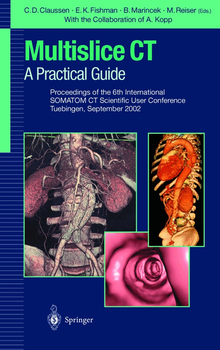
The 6th International SOMATOM CT Scientific User Conference was held in Tiibingen on September 27 and 28, 2002, under the auspices of the Eberhard Karls University Tiibingen, the University Hospital Zurich, the Ludwig Maxi milians University, Munich, and the Johns Hopkins University, Baltimore. Siemens AG Medical Solutions, Forchheim, was again very gracious in spon soring the course and the associated events. There have been remarkable achievements in CT technology, workflow management and applications since the last SOMATOM user conference in Zurich in June 2000. The next generation of multidetector-row CT, now capable of acquiring 16 slices during a single rotation has been introduced. The meeting in Tiibingen was the very first conference where research groups from allover the world were presenting their very first results with this new technology in the various fields of CT imaging. It became obvious that these new technical possi bilities will again have significant impact on the clinical use of CT. Tiibingen offered a special and inspiring atmosphere for the meeting, an atmosphere which has been preserved throughout time. For more than 500 years the university - one of the oldest in Germany - has been a source for new ideas being pursued by famous philosophers, poets and scientists such as H6lderlin, Morike, Hegel, Schelling, Kepler, Butenandt and many more.
E-bok
PDF, Engelska, 20121 408 kr
Läs direkt efter köp
Until recently, CT scanner performance was limited by a series of compromises. With single-detector scanners, one cannot select thin collimation and still maintain the required extent of volumetric coverage. Slow scans cause motion artifacts that impair image quality. The introduction of multidetector CT technology, however, has revolutionized the field. Currently multidetector, multislice CT scanners acquire up to four channels of data from interweaving spirals. The minimum gantry rotation period is as low as half of a second. This increased scan speed allows for thinner collimation and thus higher longitudinal or z-axis resolution in comparison with single-detector CT. The improved image quality with multidetector technology leads to new applications of CT, particularly in cardiac, vascular, and abdominal imaging. On-going clinical studies are evaluating the suitability of this new imaging tool for non-invasive screening and diagnosis of coronary artery disease. A particular advantage to the increased scan speed in vascular imaging is the ability to cut intra venous contrast dosage and still maintain peak enhancement CT throughout the entire acquisition. Thin-section, multiphasic acquisition during optimal arterial-phase and venous-phase enhan cement significantly improves the accuracy for small lesion and vessel detection, and enhances overall classification of abdominal neoplasms. On the other hand, the increasingly large volume data sets force to new ways of looking at, presenting, storing, and trans ferring images. Networking and two- and three dimensional data processing are the key words.

Häftad, Engelska, 2012
563 kr
Skickas inom 10-15 vardagar
The 6th International SOMATOM CT Scientific User Conference was held in Tiibingen on September 27 and 28, 2002, under the auspices of the Eberhard Karls University Tiibingen, the University Hospital Zurich, the Ludwig Maxi milians University, Munich, and the Johns Hopkins University, Baltimore. Siemens AG Medical Solutions, Forchheim, was again very gracious in spon soring the course and the associated events. There have been remarkable achievements in CT technology, workflow management and applications since the last SOMATOM user conference in Zurich in June 2000. The next generation of multidetector-row CT, now capable of acquiring 16 slices during a single rotation has been introduced. The meeting in Tiibingen was the very first conference where research groups from allover the world were presenting their very first results with this new technology in the various fields of CT imaging. It became obvious that these new technical possi bilities will again have significant impact on the clinical use of CT. Tiibingen offered a special and inspiring atmosphere for the meeting, an atmosphere which has been preserved throughout time. For more than 500 years the university - one of the oldest in Germany - has been a source for new ideas being pursued by famous philosophers, poets and scientists such as H6lderlin, Morike, Hegel, Schelling, Kepler, Butenandt and many more.
Häftad, Engelska, 2011
1 118 kr
Skickas inom 10-15 vardagar
Until recently, CT scanner performance was limited by a series of compromises. With single-detector scanners, one cannot select thin collimation and still maintain the required extent of volumetric coverage. Slow scans cause motion artifacts that impair image quality. The introduction of multidetector CT technology, however, has revolutionized the field. Currently multidetector, multislice CT scanners acquire up to four channels of data from interweaving spirals. The minimum gantry rotation period is as low as half of a second. This increased scan speed allows for thinner collimation and thus higher longitudinal or z-axis resolution in comparison with single-detector CT. The improved image quality with multidetector technology leads to new applications of CT, particularly in cardiac, vascular, and abdominal imaging. On-going clinical studies are evaluating the suitability of this new imaging tool for non-invasive screening and diagnosis of coronary artery disease. A particular advantage to the increased scan speed in vascular imaging is the ability to cut intra venous contrast dosage and still maintain peak enhancement CT throughout the entire acquisition. Thin-section, multiphasic acquisition during optimal arterial-phase and venous-phase enhan cement significantly improves the accuracy for small lesion and vessel detection, and enhances overall classification of abdominal neoplasms. On the other hand, the increasingly large volume data sets force to new ways of looking at, presenting, storing, and trans ferring images. Networking and two- and three dimensional data processing are the key words.