
Exercises in Radiological Diagnosis – serie
Visar alla böcker i serien Exercises in Radiological Diagnosis. Handla med fri frakt och snabb leverans.
10 produkter
10 produkter
Häftad, Engelska, 1982
1 116 kr
Skickas inom 10-15 vardagar
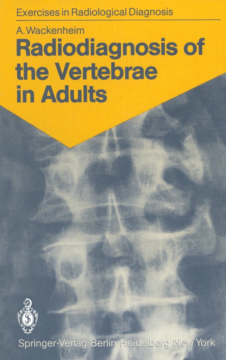
This book is intended for beginners and for those who want to refresh their knowledge of the elementary radioanatomy of the vertebrae, particularly their pathological radioanatomy. I do not pretend, as does Roger Martin du Gard's hero, that one always must begin with a radiographic examination, but I do believe that a student, especially one interested in radiology, must be able to apprehend an image isolated from its clinical context. To optimize memorization of the image I have selected unmistakable cases with marked, well-evolved lesions. This will enable the studentlater to recognize less distinct images of the same kind. The first section of the book is exclusively iconographic. After studying an image, the reader will find in the second section, under the appropriate reference number, a commen tary illustrated with a realistic drawing by my friend Dr. Csaba Hethalmi. Attention to the following points will assist a fruitful reading: 1. Cases 1-5 involve normal subjects; all other cases are pathological. 2. The reader must imagine that he is conducting a routine examination and draw on his resources to make a practical analysis of an image. As a matter of fact, all the films (except that in case 46, which is the radiograph of a specimen) were -indeed taken under routine conditions using standard pro jections. 3. The cases are in no systematic or nosological order. Each case is illustrated with one, two, or (rarely) three images.
Häftad, Engelska, 1985
1 116 kr
Skickas inom 10-15 vardagar
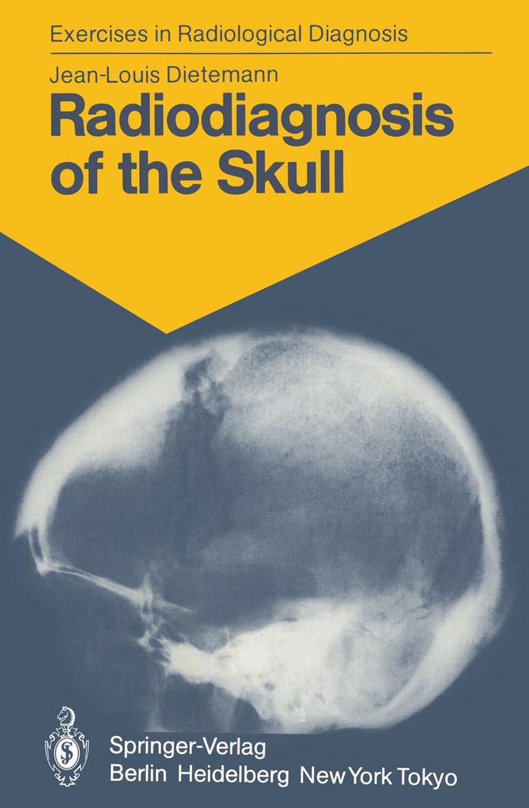
This volume is the latest in the series Exercises in Radio logical Diagnosis, launched by the Strasbourg School of Radio logy. Further volumes on the facial bones, senology, and abdominal tomography are in preparation. Jean-Louis Dietemann has over a decade's experience of radiodiagnosis of the skull, and has already proven his talents as a teacher with his earlier books on the sella turcica and on carotid angiography. The present volume will assuredly be a great success and will enhance the series. Professor A. W ACKENHEIM Hospices Civils de Strasbourg Centre Hospitalier Regional Service de Radiologie I Strasbourg v Contents Part One: Iconography . . . . . . . . . . . . .. . . 1 . Part Two: Commentary with Corresponding Schemata. 85 References . . 165 Subject Index 167 VII Part One Iconography 1 1 a b 3 2 a b 4 3 4 5 6 6 a b 7 7 8 8 a b 9 9 10 10 11 a b 11 12 13 a b 13 a b 14 b 15 16 17 16 17 20 18 21 a b 19 22 a b 20 21 24 a b 22 b 23 26 27 24 a 25 26 29 27 31 32 28 33 34 29 35 36 30 37 a b 31 38 39 32 40 41 33 42 43 34 44 a b 35 44 c 45 a 36 45 b ~----~------~~ c 37 46 47 38 48 49 39 50 a b 40 41 52 53 a 42 53 b 54 a 43 54 b 55 44 56
Häftad, Engelska, 1985
1 116 kr
Skickas inom 10-15 vardagar
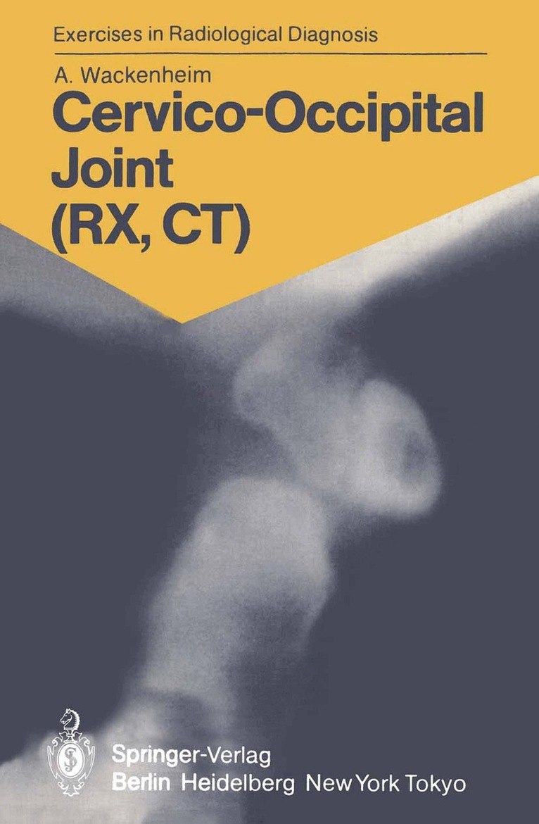
Like my first book of exercises in radiological diagnosis con- cerning the vertebrae in adults, the first part of this book consists of an atlas of images ready for immanent reading. The images are numbered, each number corresponding to a case. The second part comprises the corresponding comments, critical interpretations, and drawings (using the same num- bers as the corresponding radiographs in the first part). I hope that this second booklet will be as appreciated as was the first, which was published in four languages (Radio- diagnosis of the Vertebrae in Adults. Springer, Berlin Hei- delberg New York 1983). My own documentation having proved deficient in some fields, I had recourse to the didactic collections oimy collea- gues, friends, and students. Special thanks are due to Prof. J. F. Bonneville, and to Drs. J. L. Dietemann, Y. Dirheimer, J. C. Dosch, and J. Vignaud. AUGUSTE WACKENHEIM VII Contents Introduction ...1 Part One: Iconography 5 Part Two: Commentary with Corresponding Schemata. 107 References . .189 Subject Index 191 IX Introduction The analysis or the reading of an X-ray picture proceeds from struc- turalistic rules, such as the immanence, the synchronism, the significa- tion (signifiant and signifie of a sign), first from the semeiologic and then from the semantic point of view. In a simpler way, one can say that it is necessary to distinguish the signifiant, the signiGBPie, the comment, the interpretation, and the radiobioclinical confrontation. The signifiant or character is a normal or pathological "characteris- tic" part of the image.
Häftad, Engelska, 1988
561 kr
Skickas inom 10-15 vardagar
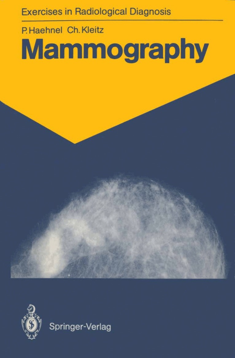
Technical advances in the field of mammography and better interpretation of the photographs have led recently to a more precise and reliable means of diagnosis, especially for findings which are difficult to analyse. With the help of numerous pathological and control cases the authors explain both the step-by-step procedure in diagnosis and the advantages of mammography as the choice of examination in particular patients. This book guides the reader to an exact interpretation of mammograms. The book is directed at radiologists, gynaecologists and general practitioners. Interdisciplinary collaboration and diagnosis in the preclinical stage are especially important for furthering the prevention of breast cancer.
Häftad, Engelska, 1986
1 116 kr
Skickas inom 10-15 vardagar
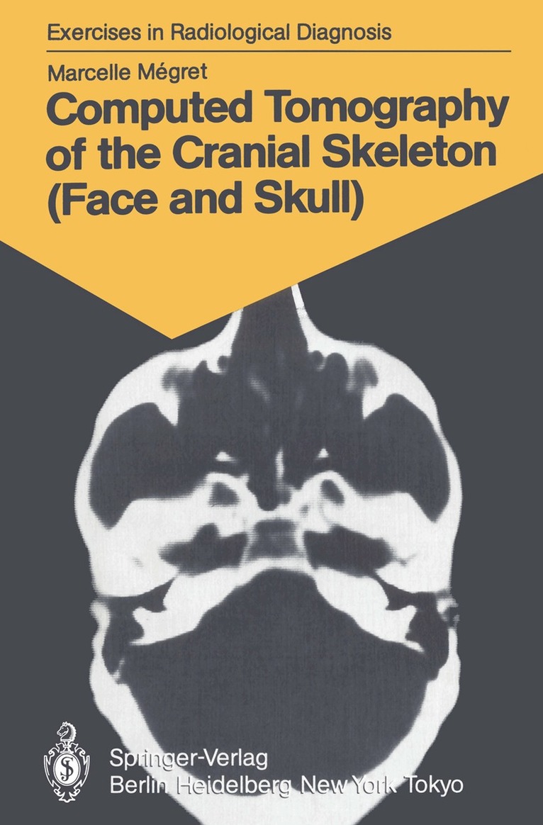
Marcelle Megret has assembled a comprehensive collection of radiographie images during her work in the field of com puted tomography of the cranial skeleton. This collection and her experience have made it possible for her to prepare these exercises in radiological diagnosis concerned exclusively with craniofacial tomodensitometry. The viewer will appreciate the choice of original docu ments of the highest quality, and also note the delicacy of execution of the diagrams, all drawn by the author herself. Marcelle Megret, like most of my students, conceives and carries out the schematas herself. The requirements concern ing certainty, strictness, precision, and objectivity would lead her to agree with Ludwig Wittgenstein's observations that since certain things cannot be said they must be shown, and that rules are insufficient when there are no examples. I should like this book to be successful. It inaugurates Marcelle Megret's career as an author and is an honor to the University of Geneva.
Häftad, Engelska, 1986
561 kr
Skickas inom 10-15 vardagar
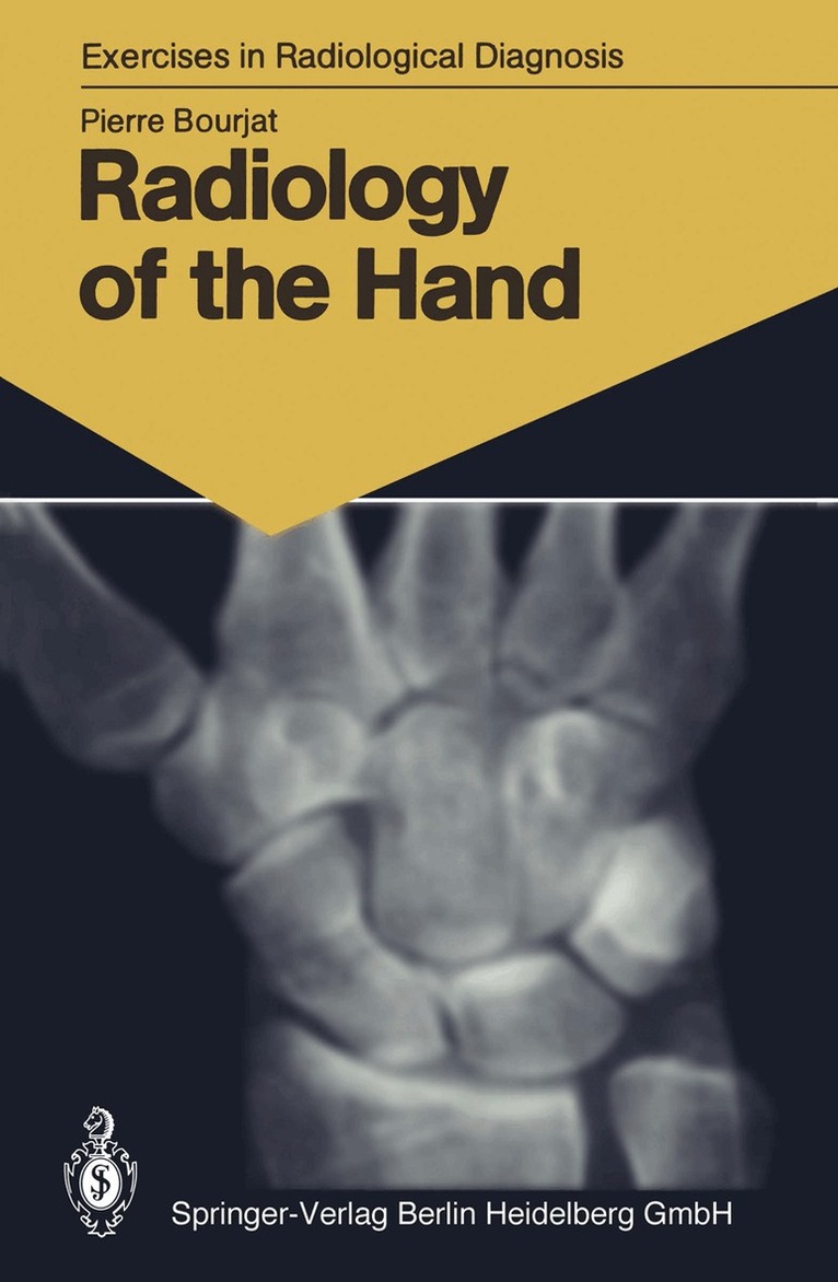
I am proud that this study of the hand appears in the series Exercises in Radiological Diagnosis founded by A. WACKEN HEIM. From time eternal, man has tried to explain the numerous configurations and lines of the hand in order to reveal the true character of a person and to display his life. The importance of the symbolism of the hand is reflected in its role in the cultural life of the old world. Therefore, it is not astanishing that the first book about the hand, by Johannes Hartlieb, was devoted to Chiromancy, and that it was so successful that it went through four editions since the second half of the fifteenth century. When Wilhelm Röntgen in 1895 asked his wife to lay her hand on a photographic plate covered with black paper and exposed it to the radiation he had just discovered, it marked the beginning of radiology. Today the study of the hand concerns only 2%-4% of the activity of radiology. It nevertheless reveals an amazing quantity and variety of pathology. That is the reason for this work. - The authors wishes to thank his friends Y. DIRHEIMER, rheumatolog1st, J. C. DoscH, traumatologist, and G. Fou CHER, surgeon, who provided some of the material for the illustrations. P. BouRJAT VII Contents Part One: Iconography . . . . . . . . . . . . . . . . 1 Part Two: Commentary with Corresponding Schemata 117 Subject Index . . . . . . . . . . . . . . . . . . 203 . . .
Computed Tomography of the Abdomen in Adults
85 Radiological Exercises for Students and Practitioners
Häftad, Engelska, 1988
561 kr
Skickas inom 10-15 vardagar
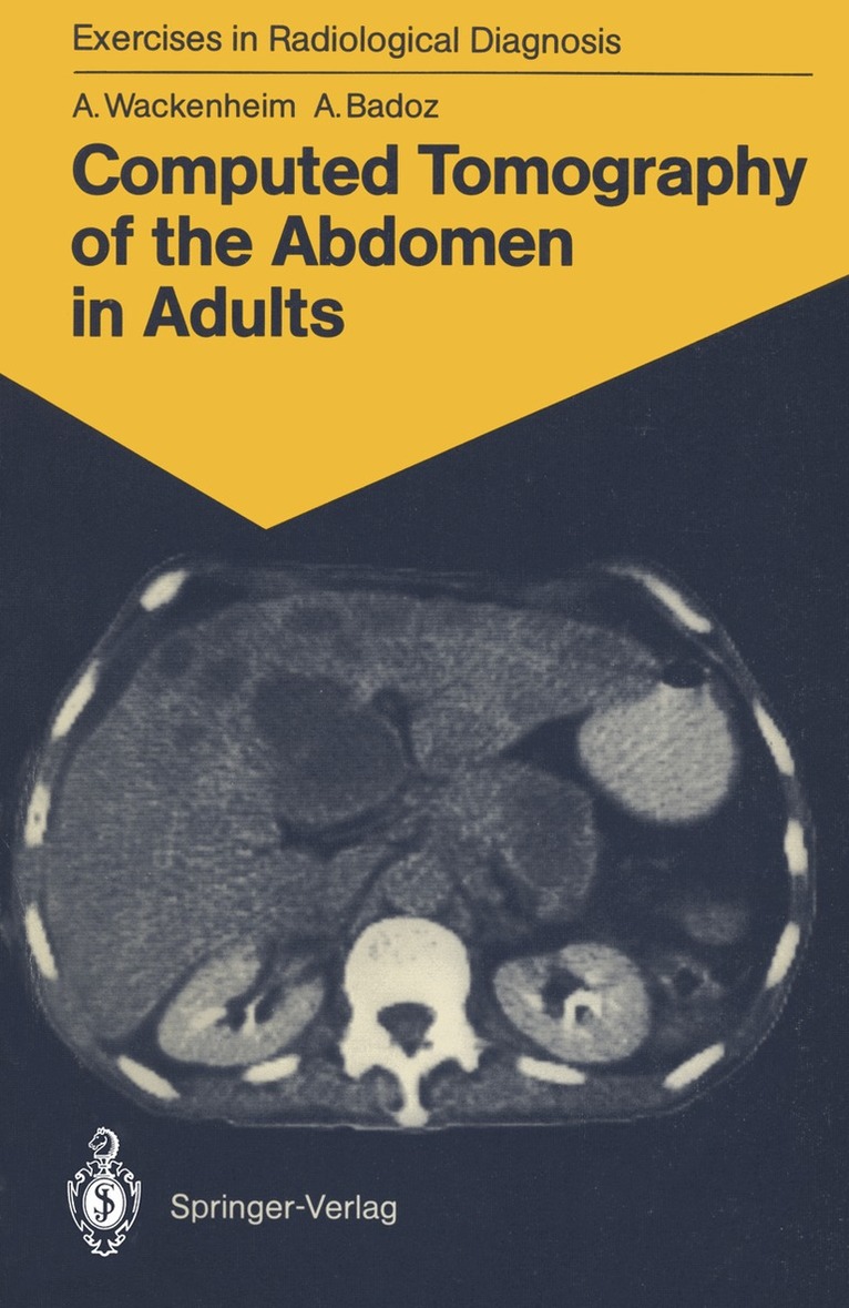
These exercises are meant for students and practitioners who wish to familiarize themselves with the normal and pathologieal computerized tomographie radioanatomy of the abdomen. The iconography is suffieiently characteristic to be read without the help of clinical or biological data. It comprises both normal and pathologie findings. Analysis of scans is comprised of two steps. The first part consists of the detailed study of normal scans, whieh serve as a reference. For this, eight main slice levels have been considered necessary and sufficient: neces sary since a certain number of slices are indispensable for the exploration of the abdomen; sufficient because a larger number of slices would risk rendering memorization difficult. The second part involves a study of the pathologie findings, organ by organ. Acknowledgements. Appreciation is extended to all those who have helped in realizing this study and, more particularly, to our friends and colleagues, J. L. DIETEMANN, C. Roy, J. L. BURGUET, M. VOUGE, and J. W. SOUITER. We would also like to thank Dr. J. WIECZOREK for his friendly assistance and advice in the planning and presentation of figures and schemata. 1 Technical Note Computerized tomography of the abdomen begins with an initial image called "scout view". This numbered radio graph of the abdomen is an analogous representation of the information and allows the location of the eight selected slice levels; these are represented by horizontallines. The slices are 10 mm thick and are taken at intervals of 2.5 cm.
Häftad, Engelska, 1987
561 kr
Skickas inom 10-15 vardagar
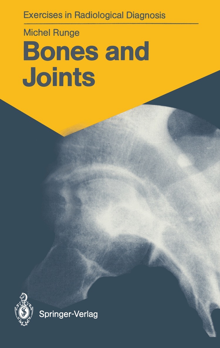
Osteoarticular pathology is a very frequent motive for consultation. Very often, the diagnosis relies upon symptomatology, and the physi cian requires confirmatory radiological investigations. Whatever the clinical indication, the interpretation of radiological data must be very rigorous. On the basis of a complete description of the radiographic images, according to a systematic analysis plan, a certain number of diagnostic hypotheses may be proposed. Selection of the most likely hypothesis requires the correlation of clinical, biological, and radiological data, and may sometimes necessi tate additional investigations, such as tomograms, scintigrams, and computed tomography (CT). 1 Part One Iconography 3 3 1 2 4 5 5 6 6 7 8 b a 8 a 9 10 11 12 10 13 14 11 15 a b 12 a c 13 17 b a c 14 15 c 16 17 23 21 a 22 b 18 19 20 21 22 23 33 34 24 25 37 38 26 27 40- 43 28 29 46 30 48 47 31 49 50 32 33 52 a b c 34 53 a b d c 35 54 a b 36 37 55 a 38 55 b c 39 56 57 40 58 41 60 61 42 43 63 64 44 65 66 45 67 68 46 69 a b 47 70 71 48 73 49 74 75 50 76 77 51 78 79 52 80 a b c 53 81 82 54 83 84 55 85 86 S6 87 88 57 89 90 58 91 92
Häftad, Engelska, 1988
561 kr
Skickas inom 10-15 vardagar
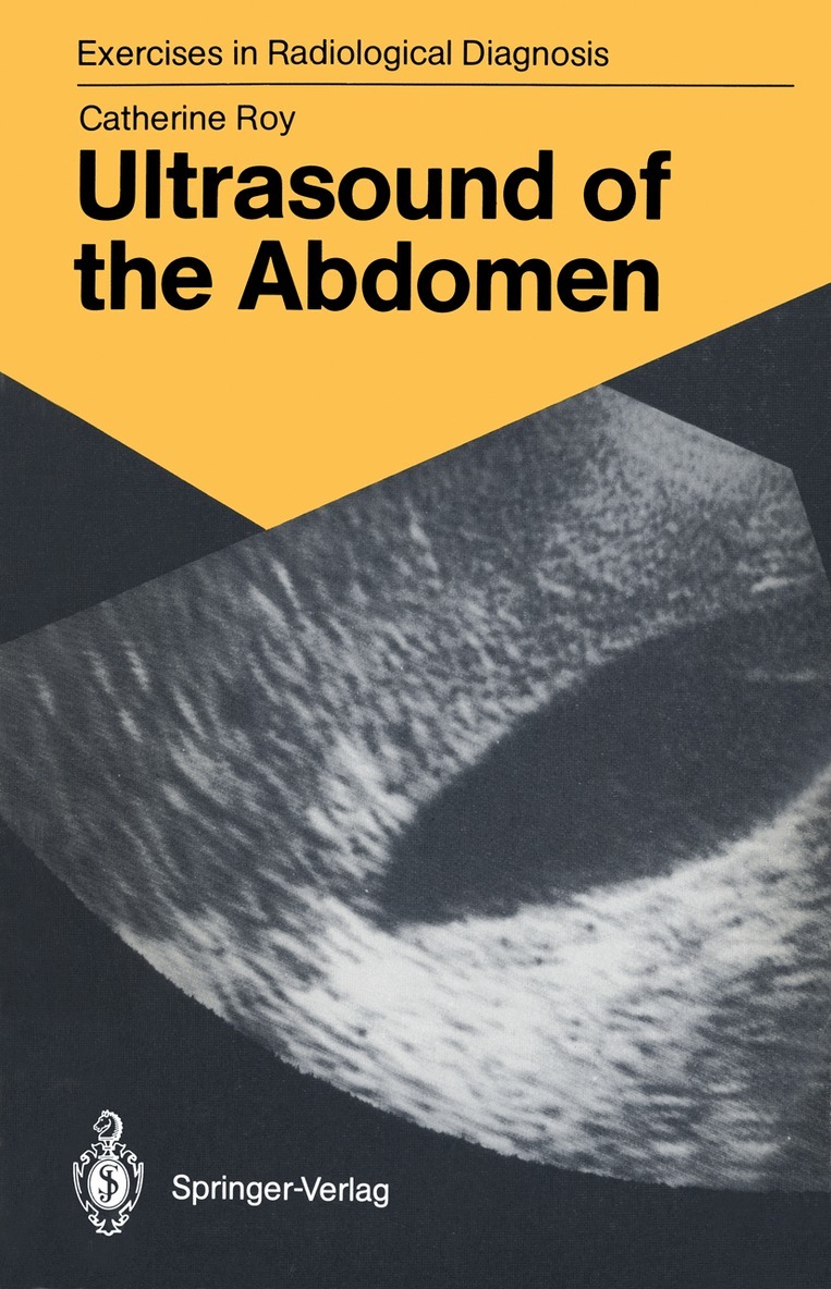
This book, the seventh in the series Exercises in Radiological Diagnosis, deals with sonography, an imaging procedure in which the ability of the radiologist plays an exceptionally important role. The author, Catherine Roy, has very extensive experience in the clinical use of sonography. She has selected the images, which are of excellent quality, with great care to illustrate a wide range of conditions and has supplemented them by commentaries and discussions which are easy to comprehend. The systematic use of schematic drawings to interpret the images makes it possible for the reader to follow the author's approach without any difficulty. Schematic drawings are par ticularly important in sonography because the relationship be tween the details in the images and the anatomy may be very weak. Images, schematic drawings, and text (both commentaries and interpretations) are three didactic elements which Cathe rine Roy has skill-fully combined in these exercises into an excellent whole. A. WACKENHEIM v Contents Introduction . . . . . . . . . . . . . . . . . . 1 Iconography, Commentary with Corresponding Schemata. . 2 Subject Index 203 VII Introduction Ultrasound imaging of the abdomen has now become a routine investi gation. It has brought about changes in the procedure of additional investigations and even rendered part of conventional radiology redun dant, particularly that concerning the bile ducts. These exercises are meant for students or physicians who already have basic knowledge of ultrasound diagnosis. It has not been possible to cover the entire spectrum of abdominal pathology, especially trauma, with 114 cases.
Häftad, Engelska, 1990
561 kr
Skickas inom 10-15 vardagar
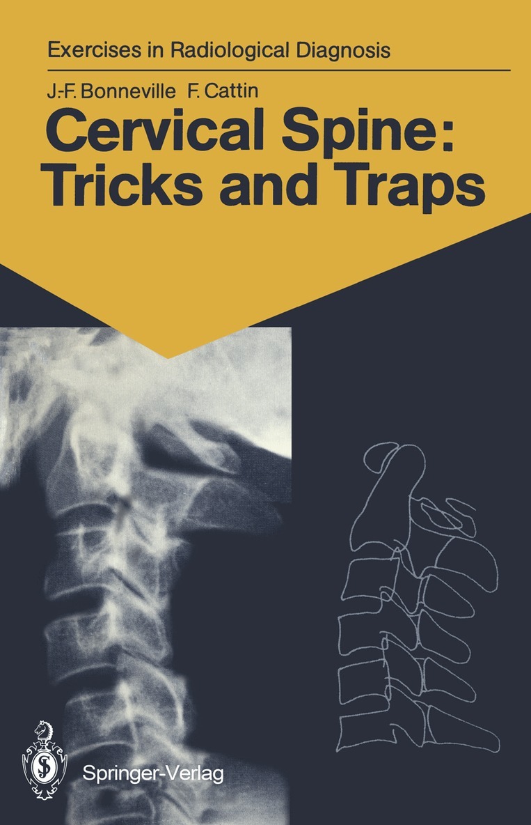
The cervical spine is always examined initially using standard radiographs, which often provide a sufficient basis for diagno- sis. Malformations, tumours, more frequently trauma, rheuma- tism, and even plain neck pain require a radiological exam- ination. The interpretation of radiological images is often difficult because of overlapping pieces of bone, the summation phe- nomena and the diversity of projections. In this book, two- or three-dimensional CTscans accompany the standard radiographs, serving as an excellent aid for com- prehension. It is almost as if the reader actually had the bones shown in the radiographs in his hands. From then on, everything becomes easy, superimpositions vanish, traps come to light, anatomy triumphs, and the images takes on life. Besanl$on, 1990 J .-F. BONNEVILLE, F. CATTIN v Contents Introduction 1 Iconography and Text with Corresponding Schemes 2 Subject Index ...123 VII Normal cervical spine: 3-D imaging 1 1 2 Case 1 1 Our starting point is a normal lateral radiograph of the cervical spine. It will serve as a guide and point of reference in our task of comprehending the interrelationships between the structures of the cervical spine.This radiograph correctly includes the entire area from the base of the skull to the cervicothora- cic junction. The soft tissues are clearly visible anteriorly, as are the extremities of the spinous processes posteriorly. In this case, with the subject looking straight ahead, is a slight, regular, physiological lordosis ofthe cervical column.